• 文献检索
  • 文档翻译
  • 深度研究
  • 学术资讯
  • Suppr Zotero 插件Zotero 插件
  • 邀请有礼
  • 套餐&价格
  • 历史记录
应用&插件
Suppr Zotero 插件Zotero 插件浏览器插件Mac 客户端Windows 客户端微信小程序
定价
高级版会员购买积分包购买API积分包
服务
文献检索文档翻译深度研究API 文档MCP 服务
关于我们
关于 Suppr公司介绍联系我们用户协议隐私条款
关注我们

Suppr 超能文献

核心技术专利:CN118964589B侵权必究
粤ICP备2023148730 号-1Suppr @ 2026

文献检索

告别复杂PubMed语法,用中文像聊天一样搜索,搜遍4000万医学文献。AI智能推荐,让科研检索更轻松。

立即免费搜索

文件翻译

保留排版,准确专业,支持PDF/Word/PPT等文件格式,支持 12+语言互译。

免费翻译文档

深度研究

AI帮你快速写综述,25分钟生成高质量综述,智能提取关键信息,辅助科研写作。

立即免费体验

磁共振磁敏感加权成像与其他成像方式在检测脾脏铁质沉着病变中的比较。

Magnetic resonance susceptibility-weighted imaging versus other imaging modalities in detecting splenic siderotic lesions.

机构信息

Department of Radiology, Southwest Hospital, Third Military Medical University, Chongqing, China.

出版信息

PLoS One. 2013 Sep 9;8(9):e73626. doi: 10.1371/journal.pone.0073626. eCollection 2013.

DOI:10.1371/journal.pone.0073626
PMID:24040004
原文链接:https://pmc.ncbi.nlm.nih.gov/articles/PMC3767753/
Abstract

BACKGROUND

Susceptibility-weighted imaging (SWI) has been proven to be superior to T2*-weighted imaging and also other existing magnetic resonance imaging (MRI) techniques for the detection of iron content and hemorrhage in the brain. The purpose of this study was to compare SWI with T1WI, T2WI and T2*WI in detecting splenic siderotic lesions.

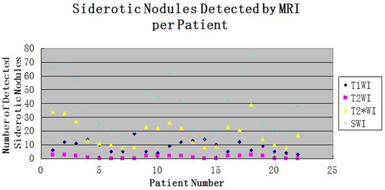
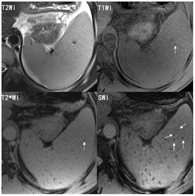
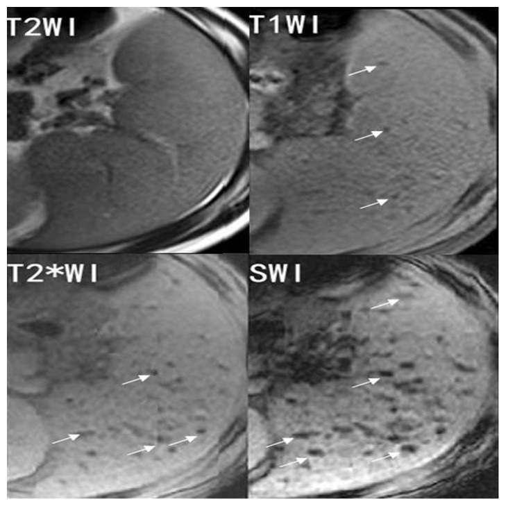
METHODOLOGY/PRINCIPAL FINDINGS: Twenty-two patients with splenic siderotic nodule were imaged with non-contrast MRI T1WI, T2WI, T2WI and SWI at 3.0 Tesla. Imaging data were independently analyzed by two experienced radiologists. The number of splenic siderotic nodules was counted, and the size (largest diameter) was measured. The conspicuity was calculated as the nodule to background parenchyma intensity ratio. We found that SWI detected a larger average number of splenic siderotic nodules than T1WI, T2WI, or T2WI (all P<0.05). The average size of the nodules detected by SWI was larger than that of those detected by T1WI, T2WI or T2*WI (all P<0.05). SWI provided superior contrast and visibility for splenic siderotic nodules compared to any other sequence (all P<0.001).

CONCLUSIONS

SWI may be a better detection scheme for splenic siderotic nodules than T1WI, T2WI and T2*WI.

摘要

背景

顺磁加权成像(SWI)已被证明在检测脑内铁含量和出血方面优于 T2*-加权成像和其他现有的磁共振成像(MRI)技术。本研究的目的是比较 SWI 与 T1WI、T2WI 和 T2*WI 在检测脾脏铁质病变中的应用。

方法/主要发现:22 例脾脏铁质结节患者在 3.0T 场强下进行非对比 MRI T1WI、T2WI、T2WI 和 SWI 扫描。两名有经验的放射科医生独立分析影像学数据。计算脾脏铁质结节的数量,并测量其大小(最大直径)。将对比噪声比作为结节与背景实质的强度比进行计算。我们发现 SWI 比 T1WI、T2WI 或 T2WI 检测到更多的脾脏铁质结节(均 P<0.05)。SWI 检测到的结节平均大小大于 T1WI、T2WI 或 T2*WI 检测到的结节(均 P<0.05)。与任何其他序列相比,SWI 为脾脏铁质结节提供了更好的对比度和可视性(均 P<0.001)。

结论

SWI 可能是一种比 T1WI、T2WI 和 T2*WI 更好的脾脏铁质结节检测方案。

https://cdn.ncbi.nlm.nih.gov/pmc/blobs/c481/3767753/79f1b22e7b5b/pone.0073626.g003.jpg
https://cdn.ncbi.nlm.nih.gov/pmc/blobs/c481/3767753/faf7baff8335/pone.0073626.g001.jpg
https://cdn.ncbi.nlm.nih.gov/pmc/blobs/c481/3767753/96bed4c07f1b/pone.0073626.g002.jpg
https://cdn.ncbi.nlm.nih.gov/pmc/blobs/c481/3767753/79f1b22e7b5b/pone.0073626.g003.jpg
https://cdn.ncbi.nlm.nih.gov/pmc/blobs/c481/3767753/faf7baff8335/pone.0073626.g001.jpg
https://cdn.ncbi.nlm.nih.gov/pmc/blobs/c481/3767753/96bed4c07f1b/pone.0073626.g002.jpg
https://cdn.ncbi.nlm.nih.gov/pmc/blobs/c481/3767753/79f1b22e7b5b/pone.0073626.g003.jpg

相似文献

1
Magnetic resonance susceptibility-weighted imaging versus other imaging modalities in detecting splenic siderotic lesions.磁共振磁敏感加权成像与其他成像方式在检测脾脏铁质沉着病变中的比较。
PLoS One. 2013 Sep 9;8(9):e73626. doi: 10.1371/journal.pone.0073626. eCollection 2013.
2
Detection of siderotic nodules in the liver with susceptibility weighted imaging: correlations to serum ferritin, Child-Pugh grade and hyaluronic acid levels.应用磁敏感加权成像检测肝脏铁沉积结节:与血清铁蛋白、Child-Pugh 分级和透明质酸水平的相关性。
Chin Med J (Engl). 2012 Sep;125(17):3110-4.
3
Improving detection of siderotic nodules in cirrhotic liver with a multi-breath-hold susceptibility-weighted imaging technique.采用多次屏气磁共振磁敏感加权成像技术提高肝硬化肝脏铁沉积结节的检出率。
J Magn Reson Imaging. 2011 Aug;34(2):318-25. doi: 10.1002/jmri.22607.
4
Improved siderotic nodule detection in cirrhosis with susceptibility-weighted magnetic resonance imaging: a prospective study.利用磁共振磁敏感加权成像提高肝硬化铁沉积性结节的检出率:一项前瞻性研究。
PLoS One. 2012;7(5):e36454. doi: 10.1371/journal.pone.0036454. Epub 2012 May 10.
5
Characterizing hepatocellular carcinoma using multi-breath-hold two-dimensional susceptibility-weighted imaging: comparison to conventional liver MRI.使用多屏息二维磁化率加权成像对肝细胞癌进行特征描述:与常规肝脏 MRI 的比较。
Clin Radiol. 2012 Dec;67(12):e91-7. doi: 10.1016/j.crad.2012.08.015. Epub 2012 Sep 13.
6
Improving detection of siderotic nodules in patients with liver disease using 2D ESWAN technique.使用二维增强型频谱加权衰减反转恢复(ESWAN)技术改善肝病患者含铁结节的检测。
Acad Radiol. 2014 Aug;21(8):971-6. doi: 10.1016/j.acra.2014.03.008.
7
Improving Detection of Iron Deposition in Cirrhotic Liver Using Susceptibility-Weighted Imaging With Emphasis on Histopathological Correlation.利用磁敏感加权成像并着重组织病理学相关性提高肝硬化肝脏中铁沉积的检测
J Comput Assist Tomogr. 2017 Jan;41(1):18-24. doi: 10.1097/RCT.0000000000000484.
8
Magnetic resonance imaging of siderotic hepatic adenoma.
J Formos Med Assoc. 1995 Mar;94(3):138-40.
9
High signal-intensity abnormalities in susceptibility-weighted imaging for primary intracerebral hemorrhage.磁敏感加权成像上的原发性脑出血高信号异常。
Int J Neurosci. 2019 Sep;129(9):842-847. doi: 10.1080/00207454.2019.1576659. Epub 2019 Mar 1.
10
Improved sensitivity of 3.0 Tesla susceptibility-weighted imaging in detecting traumatic bleeds and its use in predicting outcomes in patients with mild traumatic brain injury.3.0 特斯拉磁敏感加权成像在检测创伤性出血方面的敏感性提高及其在预测轻度创伤性脑损伤患者预后中的应用。
Acta Radiol. 2015 Oct;56(10):1256-63. doi: 10.1177/0284185114552883. Epub 2014 Oct 15.

引用本文的文献

1
Susceptibility-Weighted Imaging Manifestations in the Brain of Wilson's Disease Patients.肝豆状核变性患者脑部的磁敏感加权成像表现
PLoS One. 2015 Apr 27;10(4):e0125100. doi: 10.1371/journal.pone.0125100. eCollection 2015.

本文引用的文献

1
Hereditary hemochromatosis: pathogenesis, diagnosis, and treatment.遗传性血色素沉着症:发病机制、诊断与治疗。
Gastroenterology. 2010 Aug;139(2):393-408, 408.e1-2. doi: 10.1053/j.gastro.2010.06.013. Epub 2010 Jun 11.
2
Susceptibility-weighted imaging: technical aspects and clinical applications, part 2.磁敏感加权成像:技术要点与临床应用,第2部分。
AJNR Am J Neuroradiol. 2009 Feb;30(2):232-52. doi: 10.3174/ajnr.A1461. Epub 2009 Jan 8.
3
Susceptibility-weighted imaging: technical aspects and clinical applications, part 1.
磁敏感加权成像:技术要点与临床应用,第1部分
AJNR Am J Neuroradiol. 2009 Jan;30(1):19-30. doi: 10.3174/ajnr.A1400. Epub 2008 Nov 27.
4
Improved survival of thalassaemia major in the UK and relation to T2* cardiovascular magnetic resonance.英国重型地中海贫血患者生存率的提高及其与T2*心血管磁共振成像的关系
J Cardiovasc Magn Reson. 2008 Sep 25;10(1):42. doi: 10.1186/1532-429X-10-42.
5
The hand arthropathy of hereditary hemochromatosis is strongly associated with iron overload.遗传性血色素沉着症的手部关节病与铁过载密切相关。
J Rheumatol. 2008 Jan;35(1):153-8.
6
Haemochromatosis.血色素沉着症
Lancet. 2007 Dec 1;370(9602):1855-60. doi: 10.1016/S0140-6736(07)61782-6.
7
Magnetic resonance imaging measurement of iron overload.铁过载的磁共振成像测量
Curr Opin Hematol. 2007 May;14(3):183-90. doi: 10.1097/MOH.0b013e3280d2b76b.
8
Chronic kidney disease and mortality risk: a systematic review.慢性肾病与死亡风险:一项系统综述
J Am Soc Nephrol. 2006 Jul;17(7):2034-47. doi: 10.1681/ASN.2005101085. Epub 2006 May 31.
9
Clinical aspects of hemochromatosis.血色素沉着症的临床方面。
Semin Liver Dis. 2005 Nov;25(4):381-91. doi: 10.1055/s-2005-923310.
10
Beta-thalassemia.β地中海贫血
N Engl J Med. 2005 Sep 15;353(11):1135-46. doi: 10.1056/NEJMra050436.