Kim Sung-Mi, Kim Yong-Gun, Park Jin-Woo, Lee Jae-Mok, Suh Jo-Young
Department of Periodontology, Kyungpook National University School of Dentistry, Daegu, Korea.
J Periodontal Implant Sci. 2013 Aug;43(4):168-76. doi: 10.5051/jpis.2013.43.4.168. Epub 2013 Aug 31.
The purpose of the current study was to examine the effect of dexamethasone (Dex) at various concentrations on the apoptosis and mineralization of human periodontal ligament (hPDL) cells.
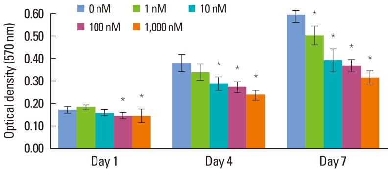
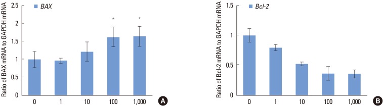
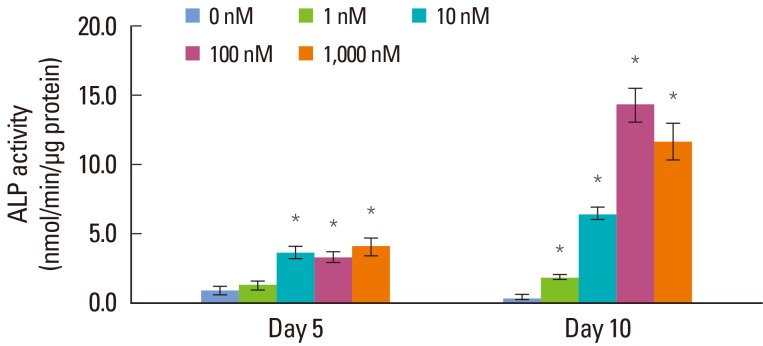
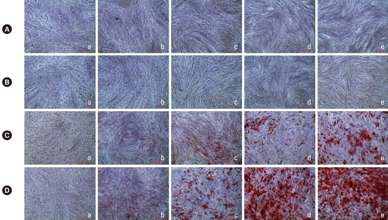
hPDL cells were obtained from the mid-third of premolars extracted for orthodontic reasons, and a primary culture of hPDL cells was prepared using an explant technique. Groups of cells were divided according to the concentration of Dex (0, 1, 10, 100, and 1,000 nM). A 3-(4,5-dimethylthiazol-2-yl)-2,5-diphenyltetrazolium bromide assay was performed for evaluation of cellular viability, and alkaline phosphatase activity was examined for osteogenic differentiation of hPDL cells. Alizarin Red S staining was performed for observation of mineralization, and real-time polymerase chain reaction was performed for the evaluation of related genes.
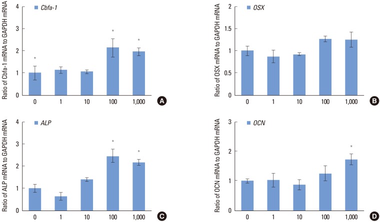
Increasing the Dex concentration was found to reduce cellular viability, with an increase in alkaline phosphatase activity and mineralization. Within the range of Dex concentrations tested in this study, 100 nM of Dex was found to promote the most vigorous differentiation and mineralization of hPDL cells. Dex-induced osteogenic differentiation and mineralization was accompanied by an increase in the level of osteogenic and apoptosis-related genes and a reduction in the level of antiapoptotic genes. The decrease in hPDL cellular viability by glucocorticoid may be explained in part by the increased prevalence of cell apoptosis, as demonstrated by BAX expression and decreased expression of the antiapoptotic gene, Bcl-2.
An increase in hPDL cell differentiation rather than cellular viability at an early stage is likely to be a key factor in glucocorticoid induced mineralization. In addition, apoptosis might play an important role in Dex-induced tissue regeneration; however, further study is needed for investigation of the precise mechanism.
本研究旨在探讨不同浓度地塞米松(Dex)对人牙周膜(hPDL)细胞凋亡和矿化的影响。
从因正畸拔除的前磨牙牙根中部三分之一处获取hPDL细胞,采用组织块培养法进行hPDL细胞的原代培养。根据Dex浓度(0、1、10、100和1000 nM)将细胞分组。采用3-(4,5-二甲基噻唑-2)-2,5-二苯基四氮唑溴盐法评估细胞活力,检测碱性磷酸酶活性以评估hPDL细胞的成骨分化情况。进行茜素红S染色观察矿化情况,采用实时聚合酶链反应评估相关基因。
发现随着Dex浓度增加,细胞活力降低,而碱性磷酸酶活性和矿化增加。在本研究测试的Dex浓度范围内,发现100 nM的Dex能促进hPDL细胞最活跃的分化和矿化。Dex诱导的成骨分化和矿化伴随着成骨及凋亡相关基因水平的升高和抗凋亡基因水平的降低。糖皮质激素导致hPDL细胞活力下降可能部分归因于细胞凋亡发生率增加,如BAX表达增加及抗凋亡基因Bcl-2表达降低所示。
早期hPDL细胞分化增加而非细胞活力增加可能是糖皮质激素诱导矿化的关键因素。此外,凋亡可能在Dex诱导的组织再生中起重要作用;然而,需要进一步研究以探讨确切机制。