Yang Xuesen, Wei Aimin, Liu Yong, He Genlin, Zhou Zhou, Yu Zhengping
Department of Occupational Health, Third Military Medical University, Chongqing, China ; Institute of Tropical Medicine, Third Military Medical University, Chongqing, China.
Mol Vis. 2013 Sep 12;19:1901-12. eCollection 2013.
Hypoxia-induced retinal ganglion cell (RGC) apoptosis has been implicated in many optic neuropathies. Insulin-like growth factor-1 (IGF-1) is important in maintaining neuronal survival, proliferation, and differentiation. The purpose of this study is to explore whether IGF-1 can protect RGCs from hypoxia-induced apoptosis and to determine the precise mechanisms that regulate this process.
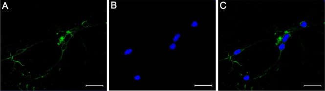
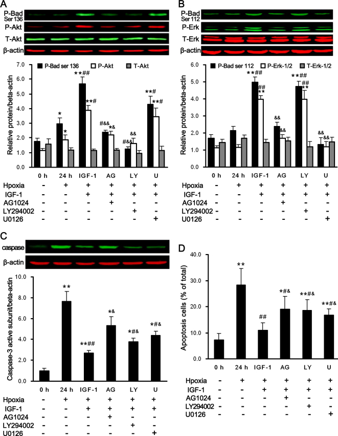
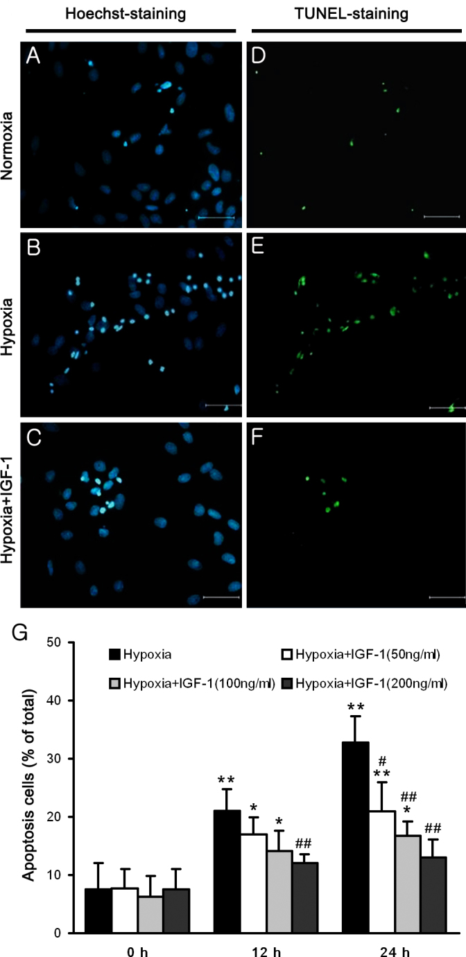
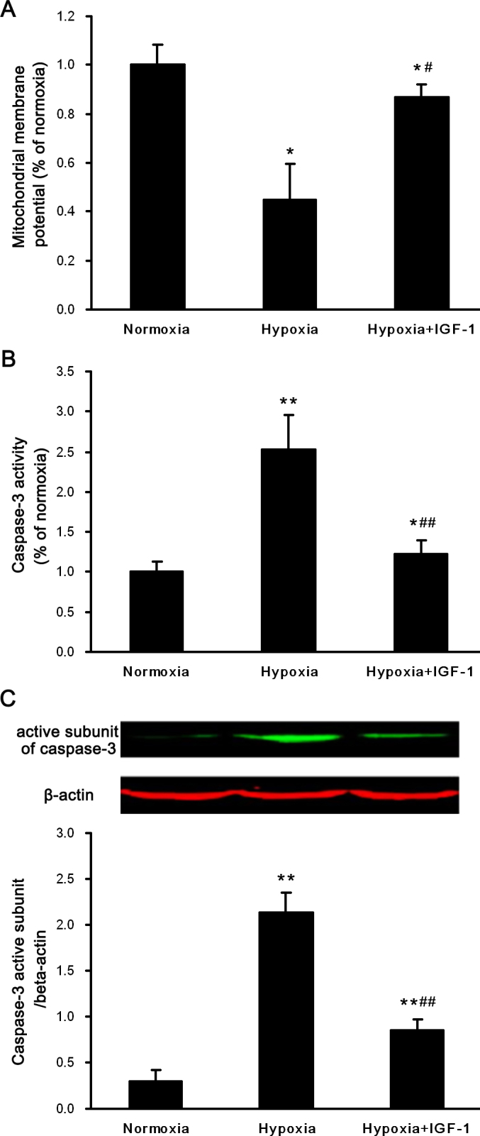
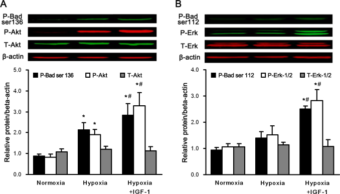
Purified RGC cultures were obtained from the retinas of neonatal Sprague Dawley (SD) rats using a two-step panning method. Primary cultured RGCs were cultured in a closed hypoxic chamber (5% O2, 5% CO2, and 90% N2) for 12 h with or without IGF-1. The degree of apoptosis in the RGCs was detected by caspase-3 expression and TUNEL and JC-1 staining assays. The expression and phosphorylation of protein kinase B (Akt), p44/42 mitogen-activated protein kinase (MAPK) (extracellular signal-regulated kinase-1/2 [Erk-1/2]), Bad, and caspase-3 was investigated with immunoblot analysis.
Hypoxia induces apoptosis in primary Sprague Dawley rat RGCs, as detected by caspase-3 expression and TUNEL and JC-1 staining assays, and that IGF-1 treatment could significantly reduce this effect in RGCs. Interestingly, pretreatment of RGCs with AG1024 (an IGF-1 inhibitor), U0126 (an Erk-1/2 inhibitor), and LY294002 (an Akt inhibitor) markedly attenuated the effects of IGF-1 treatment. Furthermore, western blot analysis suggested that the Erk-1/2 and Akt signaling pathways play a role in the protective effects of IGF-1 on RGCs exposed to hypoxia.
These data indicate that IGF-1 can protect primary cultured RGCs against hypoxia-induced apoptosis via the Erk-1/2 and Akt signaling pathways, suggesting that IGF-1 treatment is a potential therapeutic approach for treating hypoxia-induced neurodegeneration in the retina.
缺氧诱导的视网膜神经节细胞(RGC)凋亡与多种视神经病变有关。胰岛素样生长因子-1(IGF-1)在维持神经元存活、增殖和分化方面具有重要作用。本研究旨在探讨IGF-1是否能保护RGC免受缺氧诱导的凋亡,并确定调节这一过程的精确机制。
采用两步淘选法从新生Sprague Dawley(SD)大鼠的视网膜中获得纯化的RGC培养物。原代培养的RGC在封闭的缺氧培养箱(5% O2、5% CO2和90% N2)中培养12小时,分别添加或不添加IGF-1。通过半胱天冬酶-3表达、TUNEL和JC-1染色试验检测RGC中的凋亡程度。用免疫印迹分析研究蛋白激酶B(Akt)、p44/42丝裂原活化蛋白激酶(MAPK)(细胞外信号调节激酶-1/2 [Erk-1/2])、Bad和半胱天冬酶-3的表达及磷酸化情况。
通过半胱天冬酶-3表达、TUNEL和JC-1染色试验检测发现,缺氧可诱导原代Sprague Dawley大鼠RGC凋亡,而IGF-1处理可显著降低RGC中的这种效应。有趣的是,用AG1024(一种IGF-1抑制剂)、U0126(一种Erk-1/2抑制剂)和LY294002(一种Akt抑制剂)对RGC进行预处理,可明显减弱IGF-1处理的效果。此外,蛋白质印迹分析表明,Erk-1/2和Akt信号通路在IGF-1对缺氧暴露的RGC的保护作用中发挥作用。
这些数据表明,IGF-1可通过Erk-1/2和Akt信号通路保护原代培养的RGC免受缺氧诱导的凋亡,提示IGF-1治疗是治疗视网膜缺氧诱导的神经退行性变的一种潜在治疗方法。