Uzbekov Rustem, Roingeard Philippe
Plateforme des Microscopies, Université François Rabelais & CHRU de Tours, Tours, France.
BMC Res Notes. 2013 Sep 27;6:386. doi: 10.1186/1756-0500-6-386.
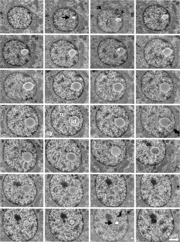
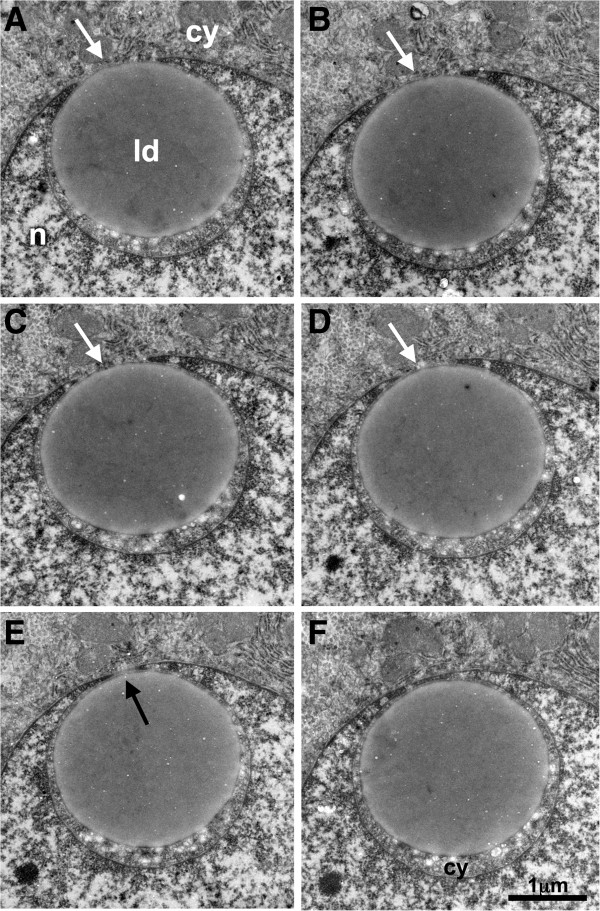
Recent studies have suggested that nuclear lipid droplets (LDs) are organized into domains similar to those of cytoplasmic LDs. As cytoplasmic LDs are formed at the endoplasmic reticulum (ER) membrane, which is structurally continuous with the nuclear envelope, it could be suggested however that nuclear LDs are cytoplamic LDs trapped within an invagination of the nuclear envelope. The resolution of fluorescence confocal microscopy is not sufficiently high to exclude this hypothesis.
We therefore addressed this question by electron microscopy (EM) of serial sections. In human liver tissue, we observed some cytoplamic LDs partly surrounded by the nuclear compartment, but we were also able to identify LDs residing in the nuclear compartment that were not connected to the nuclear envelope.
These findings indicate that nuclear LDs constitute specific subdomains of the nuclear compartment probably involved in nuclear lipid homeostasis.
最近的研究表明,核脂滴(LDs)被组织成与细胞质脂滴类似的结构域。由于细胞质脂滴在内质网(ER)膜上形成,而内质网膜与核膜在结构上是连续的,因此有人认为核脂滴可能是被困在核膜内陷处的细胞质脂滴。荧光共聚焦显微镜的分辨率不够高,无法排除这一假设。
因此,我们通过连续切片的电子显微镜(EM)来解决这个问题。在人类肝脏组织中,我们观察到一些细胞质脂滴部分被核区包围,但我们也能够识别出位于核区且与核膜无关的脂滴。
这些发现表明,核脂滴构成了核区的特定亚结构域,可能参与核脂质稳态。