Department of Genetics & Cell Biology-Molecular Biology, Cardiovascular Research Institute Maastricht (CARIM), Maastricht University, Maastricht, The Netherlands ; Institute for Molecular Cardiovascular Research (IMCAR), RWTH University Aachen, Aachen, Germany.
PLoS One. 2013 Sep 23;8(9):e75331. doi: 10.1371/journal.pone.0075331. eCollection 2013.
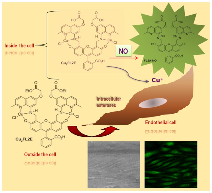
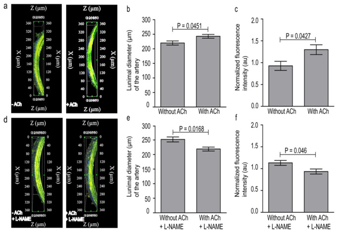
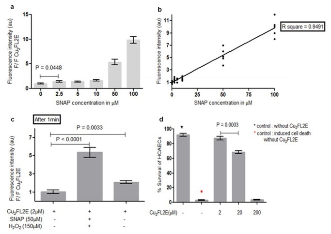
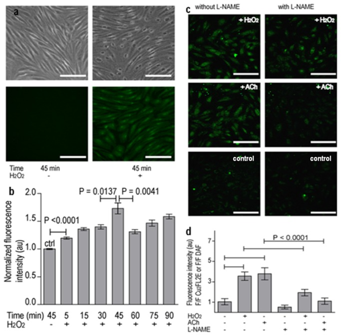
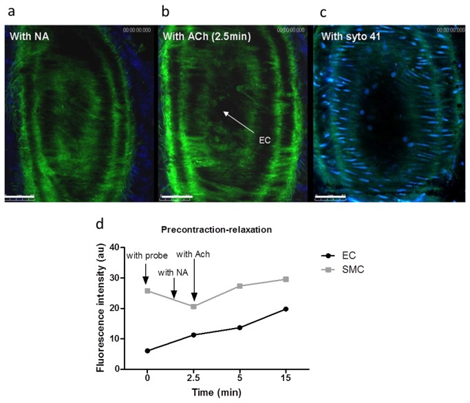
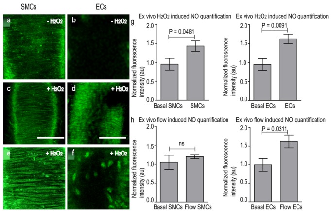
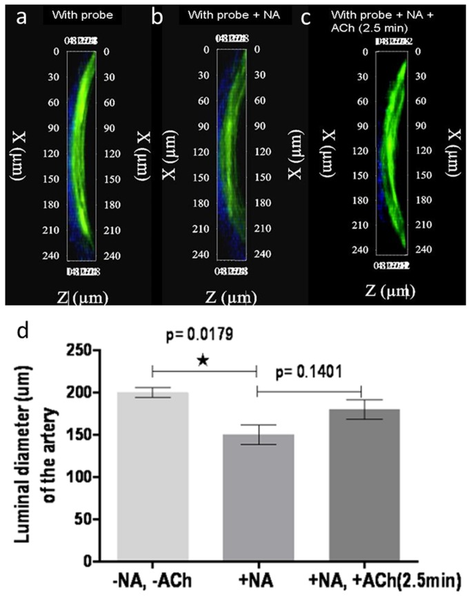
To study the role and (sub) cellular nitric oxide (NO) constitution in various disease processes, its direct and specific detection in living cells and tissues is a major requirement. Several methods are available to measure the oxidation products of NO, but the detection of NO itself has proved challenging. We visualized NO production using a NO-sensitive copper-based fluorescent probe (Cu 2FL2E) and two-photon laser scanning microscopy (TPLSM). Cu 2FL2E demonstrated high sensitivity and specificity for NO synthesis, combined with low cytotoxicity. Furthermore, Cu 2FL2E showed superior sensitivity over the conventionally used Griess assay. NO specificity of Cu 2FL2E was confirmed in vitro in human coronary arterial endothelial cells and porcine aortic endothelial cells using various triggers for NO production. Using TPLSM on ex vivo mounted murine carotid artery and aorta, the applicability of the probe to image NO production in both endothelial cells and smooth muscle cells was shown. NO-production and time course was detected for multiple stimuli such as flow, acetylcholine and hydrogen peroxide and its correlation with vasodilation was demonstrated. NO-specific fluorescence and vasodilation was abrogated in the presence of NO-synthesis blocker L-NAME. Finally, the influence of carotid precontraction and vasorelaxation validated the functional properties of vessels. Specific visualization of NO production in vessels with Cu 2FL2E-TPLSM provides a valid method for studying spatial-temporal synthesis of NO in vascular biology at an unprecedented level. This approach enables investigation of the pathways involved in the complex interplay between NO and vascular (dys) function.
为了研究各种疾病过程中(亚)细胞一氧化氮(NO)的作用,直接、特异性地检测活细胞和组织中的 NO 是主要要求。有几种方法可用于测量 NO 的氧化产物,但直接检测 NO 本身具有挑战性。我们使用对 NO 敏感的铜基荧光探针(Cu 2FL2E)和双光子激光扫描显微镜(TPLSM)可视化 NO 的产生。Cu 2FL2E 对 NO 合成具有高灵敏度和特异性,同时具有低细胞毒性。此外,Cu 2FL2E 的灵敏度优于传统的格里斯测定法。在人冠状动脉内皮细胞和猪主动脉内皮细胞中,使用各种产生 NO 的触发物,在体外证实了 Cu 2FL2E 的 NO 特异性。使用 TPLSM 对离体安装的小鼠颈总动脉和主动脉进行成像,表明该探针可用于对内皮细胞和平滑肌细胞中 NO 产生进行成像。检测了多种刺激物(如血流、乙酰胆碱和过氧化氢)的 NO 产生及其与血管舒张的相关性,并检测了其时间过程。在存在 NO 合成抑制剂 L-NAME 的情况下,NO 特异性荧光和血管舒张被阻断。最后,颈动脉预收缩和血管舒张的影响验证了血管的功能特性。使用 Cu 2FL2E-TPLSM 特异性可视化血管中 NO 的产生,为研究血管生物学中 NO 时空合成提供了一种有效的方法。这种方法能够研究 NO 与血管(功能障碍)功能之间复杂相互作用所涉及的途径。