Matsumura Nobuhisa, Shibata Takashi, Hori Emiko, Kamiyama Hironaga, Tani Mariko, Okamoto Soushi, Kubo Michiya, Horie Yukio, Endo Shunro, Kuroda Satoshi
Department of Neurosurgery, Stroke Center, Saiseikai Toyama Hospital.
Neurol Med Chir (Tokyo). 2014 Jun 17;54(6):497-501. doi: 10.2176/nmc.tn2012-0302. Epub 2013 Oct 7.
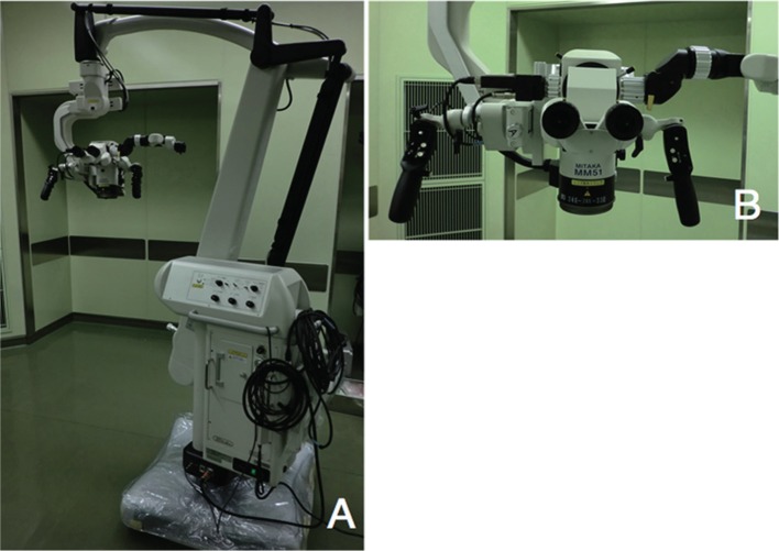
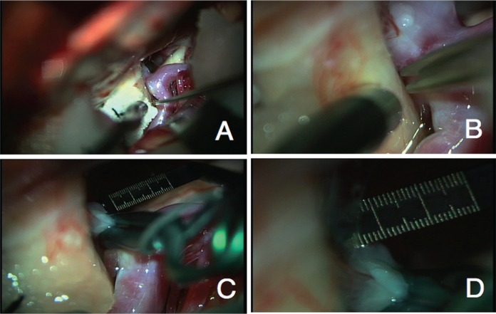
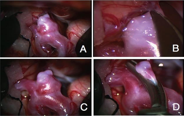
We describe a higher magnifying power operating microscope system to improve one method of high-quality microsurgical clipping for cerebral aneurysm in some cases. This higher magnification is achieved by a new lens design in the optical system, which makes the image of the object very clear at high magnifications (distinctiveness of 7 μm). This higher-resolution operating microscope system provides the surgeon with higher-magnified images (at the maximum of more than 30× magnifications as each working distance) in the operating field. The magnifications can be changed from low power (2.9×) to high power (62.0×) depending on the circumstances in a given procedure. We have used this operating microscope system on 11 patients with microsurgical clipping for cerebral aneurysms. Microsurgical treatment could be performed safely and precisely. All aneurysms were treated without any technical complications. We think that the use of this microscope would have potential benefits for microsurgical treatment for cerebral aneurysms because of better visualization.
我们描述了一种具有更高放大倍数的手术显微镜系统,在某些情况下可改进一种用于脑动脉瘤的高质量显微外科夹闭方法。这种更高的放大倍数是通过光学系统中的新透镜设计实现的,该设计使得物体在高放大倍数下(7μm 的分辨率)的图像非常清晰。这种高分辨率手术显微镜系统在手术视野中为外科医生提供更高放大倍数的图像(在每个工作距离下最大放大倍数超过 30 倍)。放大倍数可根据特定手术的情况从低倍(2.9 倍)切换到高倍(62.0 倍)。我们已将此手术显微镜系统用于 11 例接受脑动脉瘤显微外科夹闭的患者。显微外科治疗能够安全、精确地进行。所有动脉瘤治疗均无任何技术并发症。我们认为,由于可视化效果更好,使用这种显微镜对脑动脉瘤的显微外科治疗可能具有潜在益处。