Centre for Neurocognitive Research and Department of Psychology, University of Salzburg Salzburg, Austria.
Front Hum Neurosci. 2013 Sep 27;7:625. doi: 10.3389/fnhum.2013.00625. eCollection 2013.
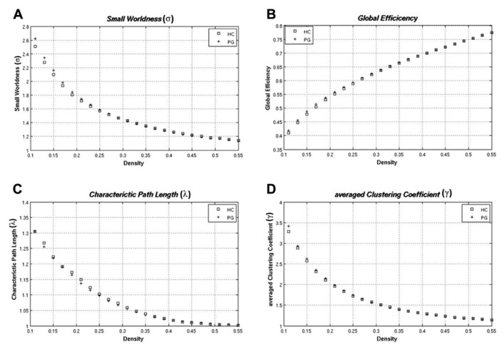
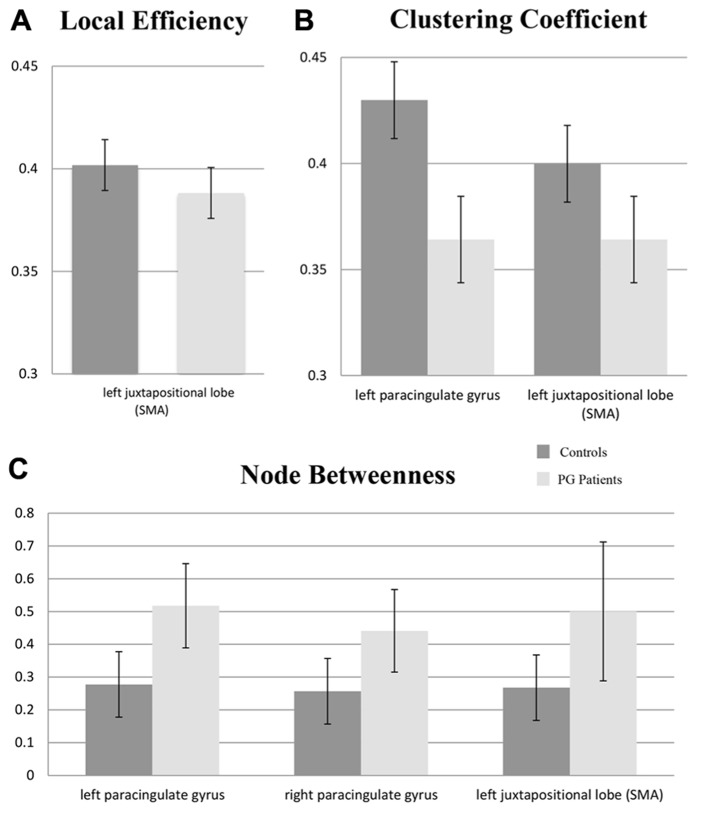
Functional neuroimaging studies of pathological gambling (PG) demonstrate alterations in frontal and subcortical regions of the mesolimbic reward system. However, most investigations were performed using tasks involving reward processing or executive functions. Little is known about brain network abnormalities during task-free resting state in PG. In the present study, graph-theoretical methods were used to investigate network properties of resting state functional magnetic resonance imaging data in PG. We compared 19 patients with PG to 19 healthy controls (HCs) using the Graph Analysis Toolbox (GAT). None of the examined global metrics differed between groups. At the nodal level, pathological gambler showed a reduced clustering coefficient in the left paracingulate cortex and the left juxtapositional lobe (supplementary motor area, SMA), reduced local efficiency in the left SMA, as well as an increased node betweenness for the left and right paracingulate cortex and the left SMA. At an uncorrected threshold level, the node betweenness in the left inferior frontal gyrus was decreased and increased in the caudate. Additionally, increased functional connectivity between fronto-striatal regions and within frontal regions has also been found for the gambling patients. These findings suggest that regions associated with the reward system demonstrate reduced segregation but enhanced integration while regions associated with executive functions demonstrate reduced integration. The present study makes evident that PG is also associated with abnormalities in the topological network structure of the brain during rest. Since alterations in PG cannot be explained by direct effects of abused substances on the brain, these findings will be of relevance for understanding functional connectivity in other addictive disorders.
功能神经影像学研究表明,病理性赌博(PG)患者的中脑边缘奖赏系统的额叶和皮质下区域发生了改变。然而,大多数研究都是使用涉及奖赏处理或执行功能的任务进行的。关于 PG 患者在无任务静息状态下的大脑网络异常,人们知之甚少。在本研究中,我们使用图论方法研究了 PG 患者静息状态功能磁共振成像数据的网络特性。我们使用 Graph Analysis Toolbox(GAT)将 19 名 PG 患者与 19 名健康对照组(HCs)进行了比较。在检查的全局指标中,两组之间没有差异。在节点水平上,病理性赌徒的左侧旁中央皮质和左侧毗邻叶(辅助运动区,SMA)的聚类系数降低,左侧 SMA 的局部效率降低,左侧和右侧旁中央皮质以及左侧 SMA 的节点介数增加。在未校正的阈值水平上,左侧额下回的节点介数降低,尾状核的节点介数增加。此外,还发现赌博患者的额纹状体区域之间以及额区内部的功能连接增加。这些发现表明,与奖赏系统相关的区域表现出降低的分离但增强的整合,而与执行功能相关的区域表现出降低的整合。本研究表明,PG 还与大脑在休息时的拓扑网络结构异常有关。由于 PG 的改变不能用滥用物质对大脑的直接影响来解释,这些发现对于理解其他成瘾障碍中的功能连接将具有重要意义。