Hales Patrick W, Phipps Kim P, Kaur Ramneek, Clark Christopher A
Imaging and Biophysics Unit, Institute of Child Health, University College London, London, United Kingdom.
PLoS One. 2013 Oct 2;8(10):e75717. doi: 10.1371/journal.pone.0075717. eCollection 2013.
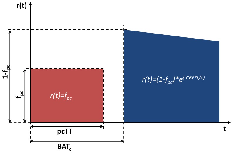
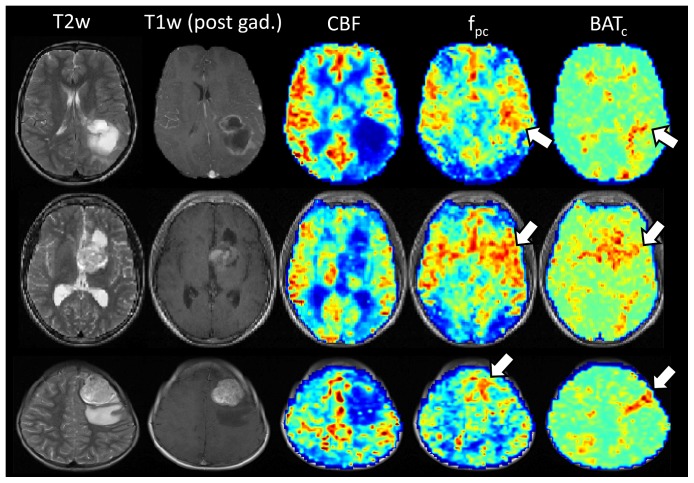
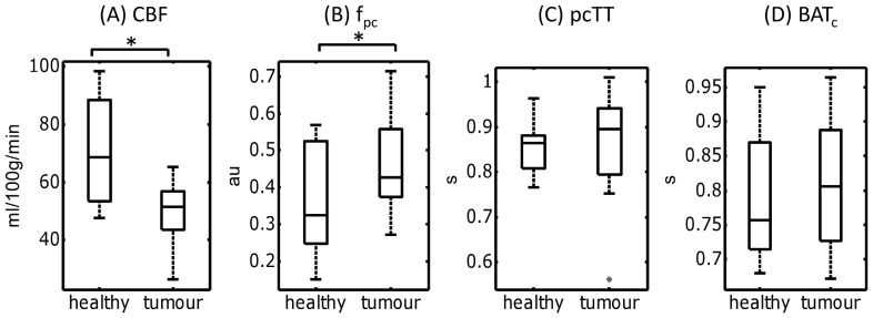
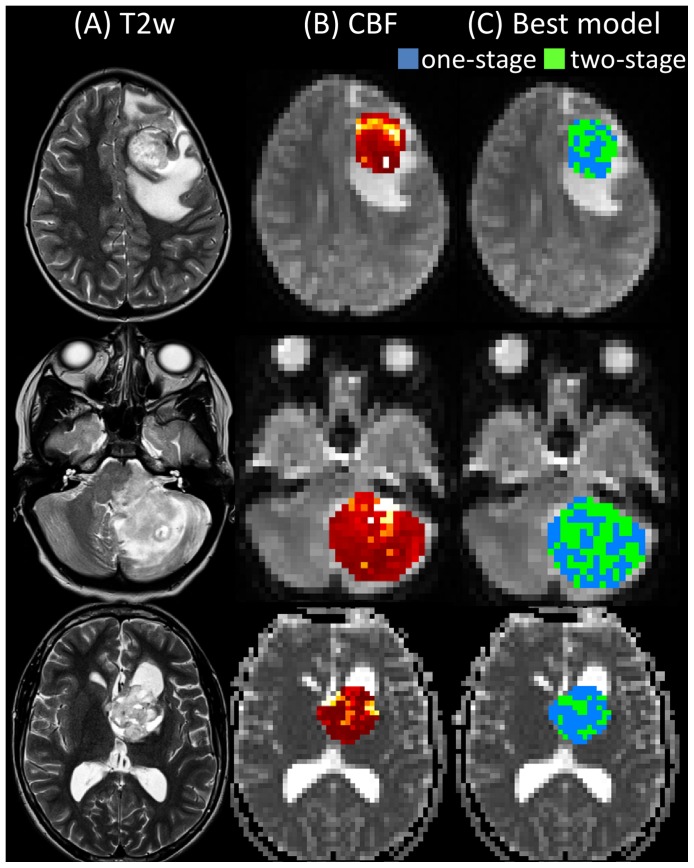
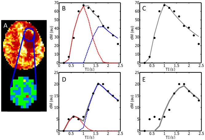
The ability to assess brain tumor perfusion and abnormalities in the vascular structure in vivo could provide significant benefits in terms of lesion diagnosis and assessment of treatment response. Arterial spin labeling (ASL) has emerged as an increasingly viable methodology for non-invasive assessment of perfusion. Although kinetic models have been developed to describe perfusion in healthy tissue, the dynamic behaviour of the ASL signal in the brain tumor environment has not been extensively studied. We show here that dynamic ASL data acquired in brain tumors displays an increased level of 'biphasic' behaviour, compared to that seen in healthy tissue. A new two-stage model is presented which more accurately describes this behaviour, and provides measurements of perfusion, pre-capillary blood volume fraction and transit time, and capillary bolus arrival time. These biomarkers offer a novel contrast in the tumor and surrounding tissue, and provide a means for measuring tumor perfusion and vascular structural abnormalities in a fully non-invasive manner.
在体内评估脑肿瘤灌注及血管结构异常的能力,在病变诊断和治疗反应评估方面可能带来显著益处。动脉自旋标记(ASL)已成为一种越来越可行的非侵入性灌注评估方法。尽管已经开发出动力学模型来描述健康组织中的灌注,但脑肿瘤环境中ASL信号的动态行为尚未得到广泛研究。我们在此表明,与健康组织相比,脑肿瘤中获取的动态ASL数据显示出更高水平的“双相”行为。提出了一种新的两阶段模型,该模型能更准确地描述这种行为,并提供灌注、毛细血管前血容量分数、通过时间以及毛细血管团到达时间的测量值。这些生物标志物在肿瘤及周围组织中提供了一种新的对比,并提供了一种以完全非侵入性方式测量肿瘤灌注和血管结构异常的方法。