Hoffmann Gabriel, Preibisch Christine, Günther Matthias, Mahroo Amnah, van Osch Matthias J P, Václavů Lena, Metz Marie-Christin, Jung Kirsten, Zimmer Claus, Wiestler Benedikt, Kaczmarz Stephan
School of Medicine and Health, Institute for Diagnostic and Interventional Neuroradiology, Technical University of Munich, Munich, Germany.
School of Medicine and Health, TUM-Neuroimaging Center, Technical University of Munich, Munich, Germany.
Magn Reson Med. 2025 May;93(5):2086-2098. doi: 10.1002/mrm.30415. Epub 2025 Jan 8.
In brain tumors, disruption of the blood-brain barrier (BBB) indicates malignancy. Clinical assessment is qualitative; quantitative evaluation is feasible using the K leakage parameter from dynamic susceptibility contrast MRI. However, contrast agent-based techniques are limited in patients with renal dysfunction and insensitive to subtle impairments. Assessing water transport times across the BBB (T) by multi-echo arterial spin labeling promises to detect BBB impairments noninvasively and potentially more sensitively. We hypothesized that reduced T indicates impaired BBB. Furthermore, we assumed higher sensitivity for T than dynamic susceptibility contrast-based K, because arterial spin labeling uses water as a freely diffusible tracer.
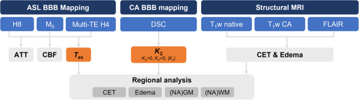
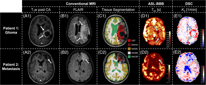
We acquired 3T MRI data from 28 patients with intraparenchymal brain tumors (World Health Organization Grade 3 & 4 gliomas [n = 17] or metastases [n = 11]) and 17 age-matched healthy controls. The protocol included multi-echo and single-echo Hadamard-encoded arterial spin labeling, dynamic susceptibility contrast, and conventional clinical imaging. T was calculated using a T-dependent multi-compartment model. Areas of contrast-enhancing tissue, edema, and normal-appearing tissue were automatically segmented, and parameter values were compared across volumes of interest and between patients and healthy controls.
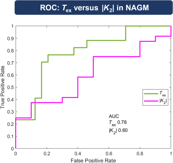
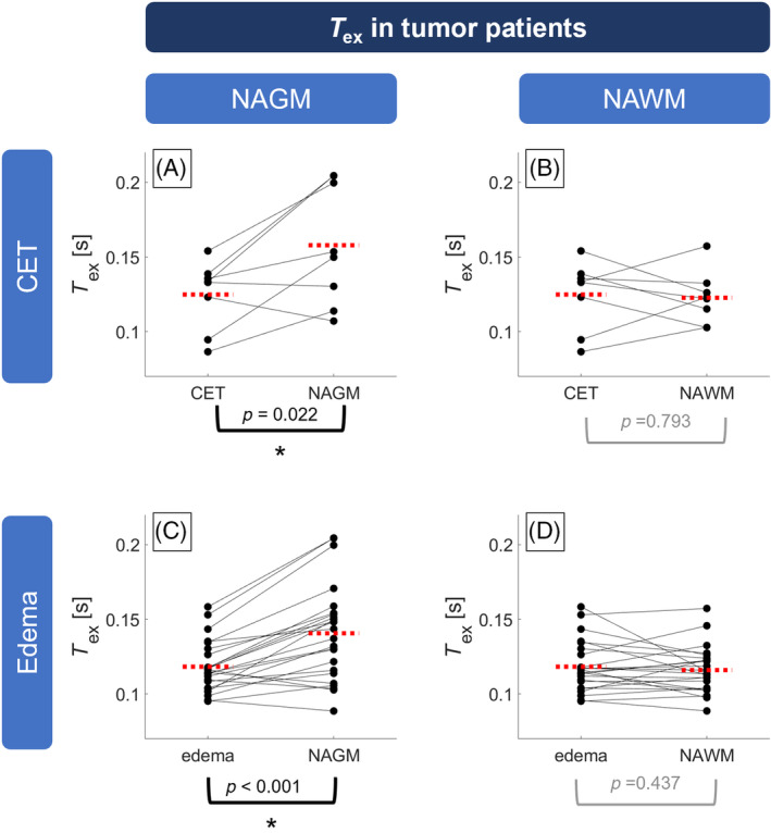
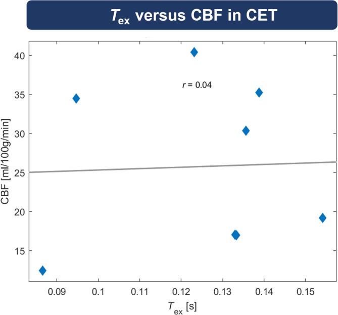
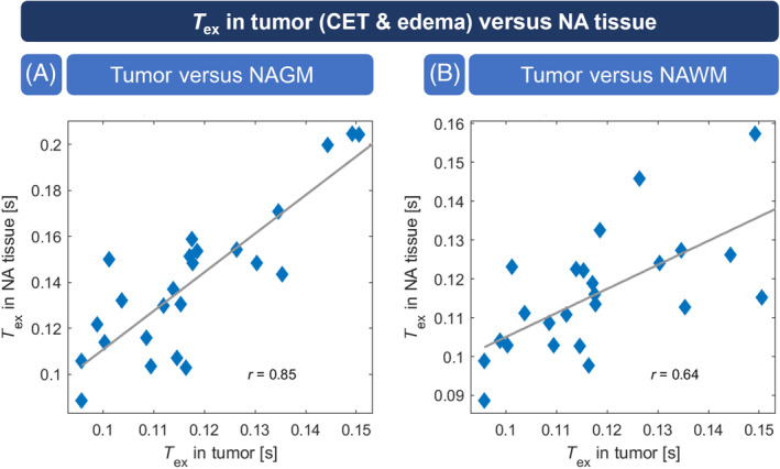
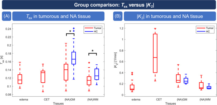
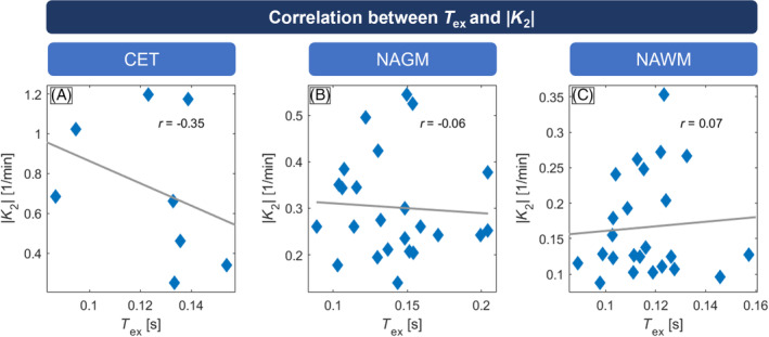
T was significantly reduced (-20.3%) in contrast-enhancing tissue compared with normal-appearing gray matter and correlated well with |K| (r = -0.347). Compared with healthy controls, T was significantly lower in tumor patients' normal-appearing gray matter (T = 0.141 ± 0.032 s vs. T = 0.172 ± 0.036 s) and normal-appearing white matter (T = 0.116 ± 0.015 vs. T = 0.127 ± 0.017 s), whereas |K| did not differ significantly. Receiver operating characteristic analysis showed a larger area under the curve for T (0.784) than K (0.604).
T is sensitive to pathophysiologically impaired BBB. It agrees with contrast agent-based K in contrast-enhancing tissue and indicates sensitivity to subtle leakage.
在脑肿瘤中,血脑屏障(BBB)的破坏表明存在恶性肿瘤。临床评估是定性的;使用动态磁敏感对比增强磁共振成像(MRI)的K泄漏参数进行定量评估是可行的。然而,基于造影剂的技术在肾功能不全患者中存在局限性,并且对细微损伤不敏感。通过多回波动脉自旋标记评估水通过血脑屏障的时间(T)有望无创且可能更敏感地检测血脑屏障损伤。我们假设T降低表明血脑屏障受损。此外,我们认为T比基于动态磁敏感对比增强的K具有更高的敏感性,因为动脉自旋标记使用水作为自由扩散的示踪剂。
我们从28例脑实质内脑肿瘤患者(世界卫生组织3级和4级胶质瘤[n = 17]或转移瘤[n = 11])和17名年龄匹配的健康对照者中获取了3T MRI数据。该方案包括多回波和单回波哈达玛编码动脉自旋标记、动态磁敏感对比增强以及传统临床成像。使用依赖于T的多室模型计算T。自动分割对比增强组织、水肿和外观正常组织的区域,并在感兴趣体积之间以及患者与健康对照者之间比较参数值。
与外观正常的灰质相比,对比增强组织中的T显著降低(-20.3%),并且与|K|具有良好的相关性(r = -0.347)。与健康对照者相比,肿瘤患者外观正常的灰质(T = 0.141±0.032秒对T = 0.172±0.036秒)和外观正常的白质(T = 0.116±0.015对T = 0.127±0.017秒)中的T显著更低,而|K|没有显著差异。受试者工作特征分析显示,T的曲线下面积(0.784)大于K(0.604)。
T对病理生理受损的血脑屏障敏感。在对比增强组织中,它与基于造影剂的K一致,并表明对细微渗漏敏感。