Hess Agathe, Yu Lianchun, Klein Isabelle, De Mazancourt Marine, Jebrak Gilles, Mal Hervé, Brugière Olivier, Fournier Michel, Courbage Maurice, Dauriat Gaelle, Schouman-Clayes Elisabeth, Clerici Christine, Mangin Laurence
Laboratoire Matière et Systèmes complexes, UMR 7057, CNRS, Université Paris 7, Paris, France ; Service de Radiologie, APHP, Hôpital Bichat-Claude Bernard, Paris, France.
PLoS One. 2013 Oct 3;8(10):e75740. doi: 10.1371/journal.pone.0075740. eCollection 2013.
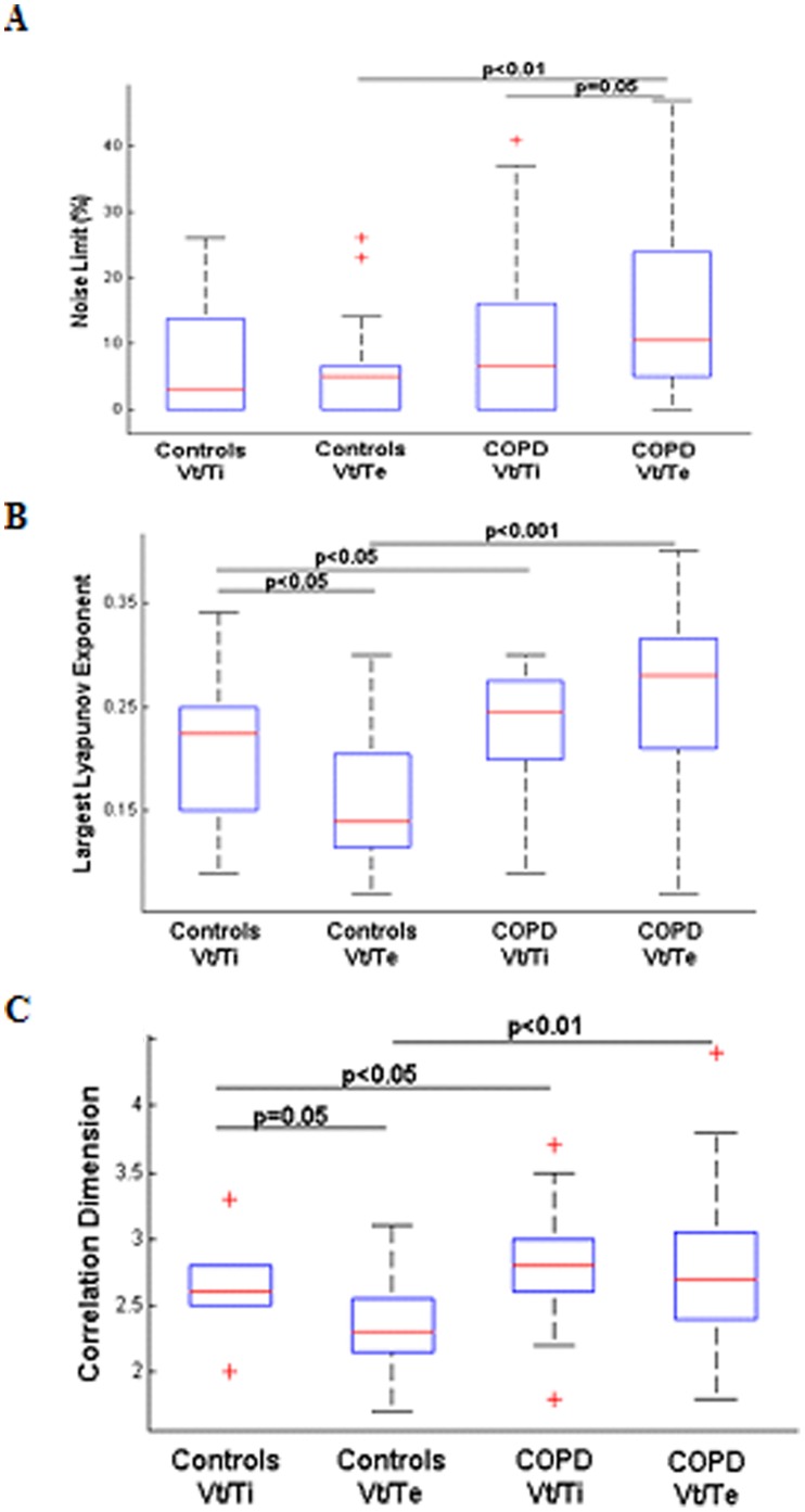
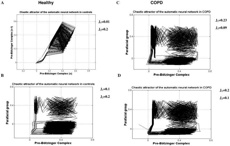
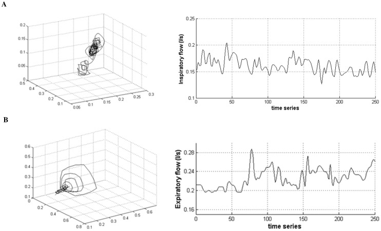
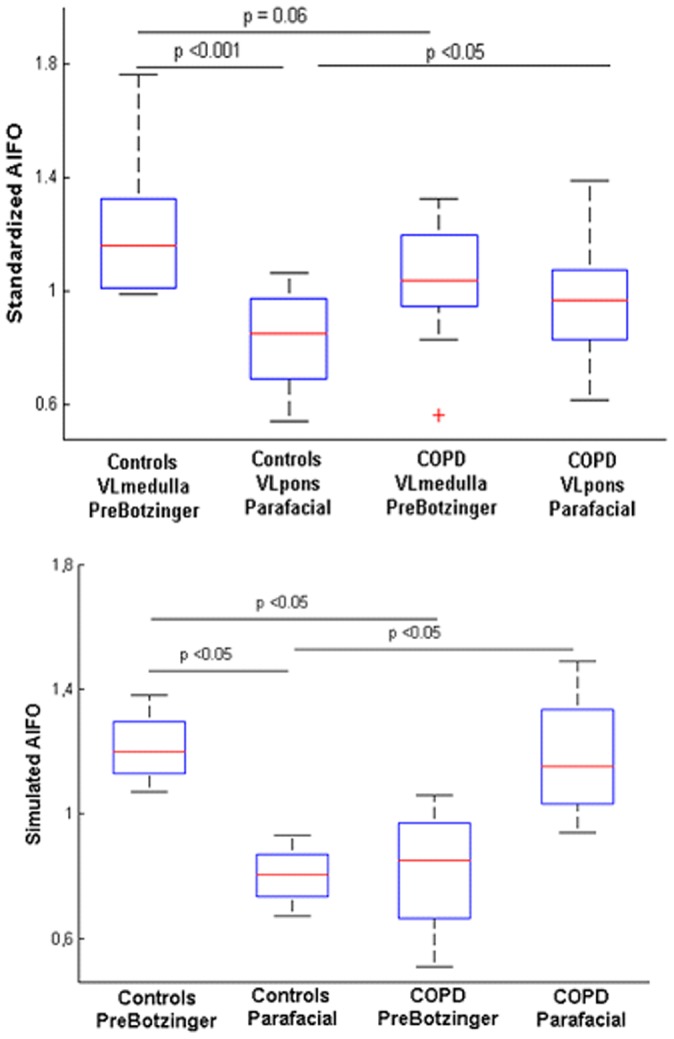
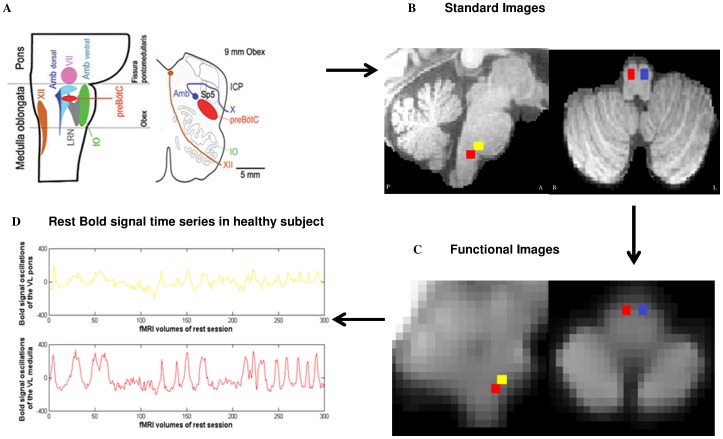
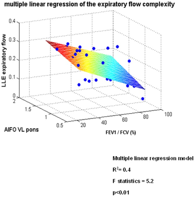
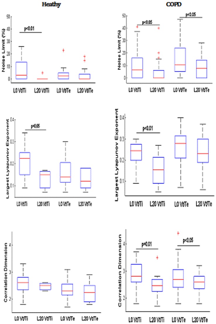
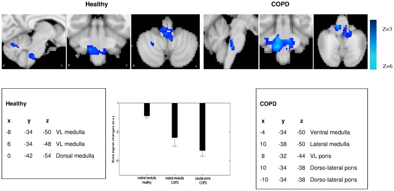
Breathing is maintained and controlled by a network of automatic neurons in the brainstem that generate respiratory rhythm and receive regulatory inputs. Breathing complexity therefore arises from respiratory central pattern generators modulated by peripheral and supra-spinal inputs. Very little is known on the brainstem neural substrates underlying breathing complexity in humans. We used both experimental and theoretical approaches to decipher these mechanisms in healthy humans and patients with chronic obstructive pulmonary disease (COPD). COPD is the most frequent chronic lung disease in the general population mainly due to tobacco smoke. In patients, airflow obstruction associated with hyperinflation and respiratory muscles weakness are key factors contributing to load-capacity imbalance and hence increased respiratory drive. Unexpectedly, we found that the patients breathed with a higher level of complexity during inspiration and expiration than controls. Using functional magnetic resonance imaging (fMRI), we scanned the brain of the participants to analyze the activity of two small regions involved in respiratory rhythmogenesis, the rostral ventro-lateral (VL) medulla (pre-Bötzinger complex) and the caudal VL pons (parafacial group). fMRI revealed in controls higher activity of the VL medulla suggesting active inspiration, while in patients higher activity of the VL pons suggesting active expiration. COPD patients reactivate the parafacial to sustain ventilation. These findings may be involved in the onset of respiratory failure when the neural network becomes overwhelmed by respiratory overload We show that central neural activity correlates with airflow complexity in healthy subjects and COPD patients, at rest and during inspiratory loading. We finally used a theoretical approach of respiratory rhythmogenesis that reproduces the kernel activity of neurons involved in the automatic breathing. The model reveals how a chaotic activity in neurons can contribute to chaos in airflow and reproduces key experimental fMRI findings.
呼吸由脑干中一组自动神经元网络维持和控制,这些神经元产生呼吸节律并接收调节输入。因此,呼吸复杂性源于受外周和脊髓上输入调节的呼吸中枢模式发生器。关于人类呼吸复杂性背后的脑干神经基质,我们所知甚少。我们采用实验和理论方法来解读健康人和慢性阻塞性肺疾病(COPD)患者的这些机制。COPD是普通人群中最常见的慢性肺病,主要由烟草烟雾引起。在患者中,与肺过度充气和呼吸肌无力相关的气流阻塞是导致负荷 - 能力失衡进而增加呼吸驱动力的关键因素。出乎意料的是,我们发现患者在吸气和呼气时的呼吸复杂性水平高于对照组。我们使用功能磁共振成像(fMRI)扫描参与者的大脑,以分析参与呼吸节律产生的两个小区域的活动,即延髓头端腹外侧(VL)(前包钦格复合体)和脑桥尾端VL(面神经旁组)。fMRI显示,在对照组中,VL延髓的活动较高,表明主动吸气,而在患者中,VL脑桥的活动较高,表明主动呼气。COPD患者重新激活面神经旁组以维持通气。当神经网络因呼吸负荷过重而不堪重负时,这些发现可能与呼吸衰竭的发生有关。我们表明,在健康受试者和COPD患者中,静息和吸气负荷期间,中枢神经活动与气流复杂性相关。我们最终采用了一种呼吸节律产生的理论方法,该方法再现了参与自动呼吸的神经元的核心活动。该模型揭示了神经元中的混沌活动如何导致气流混沌,并再现了关键的实验性fMRI结果。