Yu Lianchun, De Mazancourt Marine, Hess Agathe, Ashadi Fakhrul R, Klein Isabelle, Mal Hervé, Courbage Maurice, Mangin Laurence
Department of Physics, Matter and Complex Systems Research Laboratory, UMR 7057, CNRS, Paris 7 University, France.
Institute of Theoretical Physics, Lanzhou University, Lanzhou, China.
Hum Brain Mapp. 2016 Aug;37(8):2736-54. doi: 10.1002/hbm.23205. Epub 2016 Apr 5.
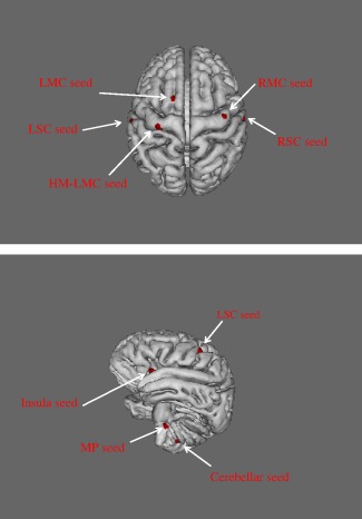
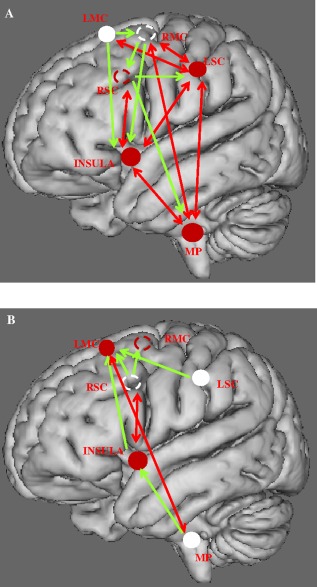
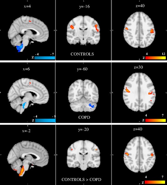
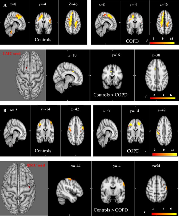
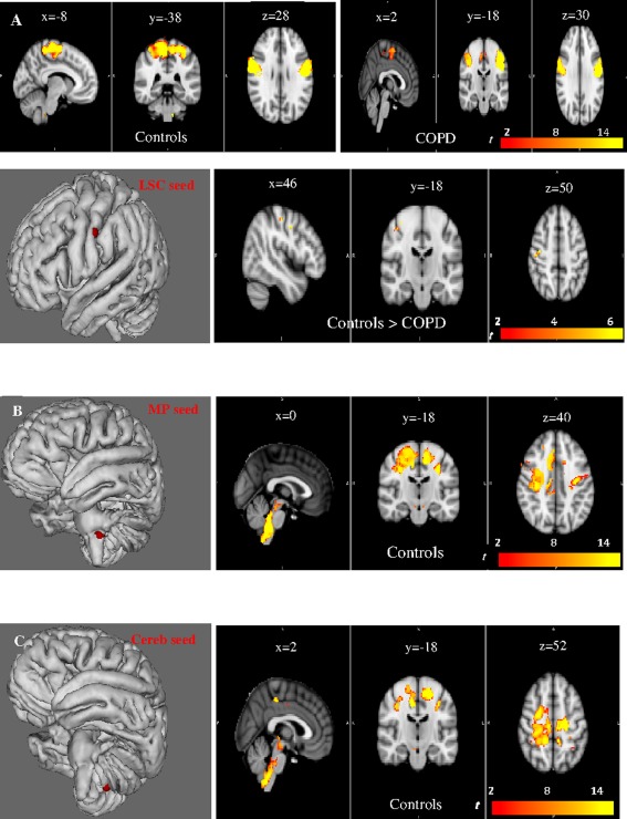
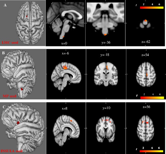
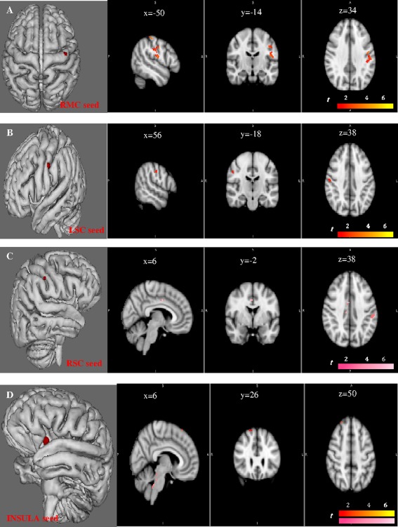
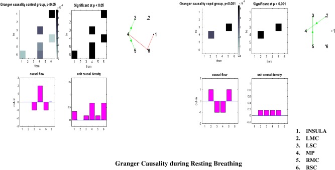
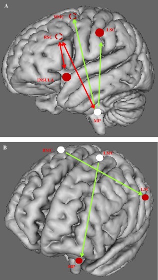
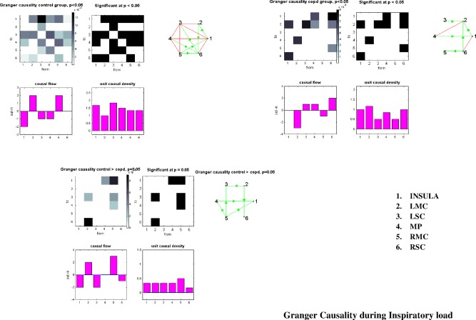
Breathing involves a complex interplay between the brainstem automatic network and cortical voluntary command. How these brain regions communicate at rest or during inspiratory loading is unknown. This issue is crucial for several reasons: (i) increased respiratory loading is a major feature of several respiratory diseases, (ii) failure of the voluntary motor and cortical sensory processing drives is among the mechanisms that precede acute respiratory failure, (iii) several cerebral structures involved in responding to inspiratory loading participate in the perception of dyspnea, a distressing symptom in many disease. We studied functional connectivity and Granger causality of the respiratory network in controls and patients with chronic obstructive pulmonary disease (COPD), at rest and during inspiratory loading. Compared with those of controls, the motor cortex area of patients exhibited decreased connectivity with their contralateral counterparts and no connectivity with the brainstem. In the patients, the information flow was reversed at rest with the source of the network shifted from the medulla towards the motor cortex. During inspiratory loading, the system was overwhelmed and the motor cortex became the sink of the network. This major finding may help to understand why some patients with COPD are prone to acute respiratory failure. Network connectivity and causality were related to lung function and illness severity. We validated our connectivity and causality results with a mathematical model of neural network. Our findings suggest a new therapeutic strategy involving the modulation of brain activity to increase motor cortex functional connectivity and improve respiratory muscles performance in patients. Hum Brain Mapp 37:2736-2754, 2016. © 2016 The Authors Human Brain Mapping Published by Wiley Periodicals, Inc.
呼吸涉及脑干自动网络与皮层自主指令之间复杂的相互作用。这些脑区在静息状态或吸气负荷期间如何进行通信尚不清楚。由于以下几个原因,这个问题至关重要:(i)呼吸负荷增加是几种呼吸系统疾病的主要特征;(ii)自主运动和皮层感觉处理驱动功能的衰竭是急性呼吸衰竭之前的机制之一;(iii)参与对吸气负荷作出反应的几个脑结构参与了呼吸困难的感知,呼吸困难是许多疾病中令人痛苦的症状。我们研究了对照组和慢性阻塞性肺疾病(COPD)患者在静息状态和吸气负荷期间呼吸网络的功能连接性和格兰杰因果关系。与对照组相比,COPD患者的运动皮层区域与其对侧对应区域的连接性降低,且与脑干无连接。在COPD患者中,静息时信息流发生逆转,网络源从延髓转移至运动皮层。在吸气负荷期间,该系统不堪重负,运动皮层成为网络的汇聚点。这一主要发现可能有助于理解为什么一些COPD患者易发生急性呼吸衰竭。网络连接性和因果关系与肺功能及疾病严重程度相关。我们用神经网络数学模型验证了我们的连接性和因果关系结果。我们的研究结果提示了一种新的治疗策略,即通过调节脑活动来增加运动皮层功能连接性,并改善患者呼吸肌的性能。《人类脑图谱》37:2736 - 2754,2016年。© 2016作者。《人类脑图谱》由威利期刊公司出版。