Max F. Perutz Laboratories, Medical University of Vienna, Vienna, Austria.
PLoS One. 2013 Oct 3;8(10):e76715. doi: 10.1371/journal.pone.0076715. eCollection 2013.
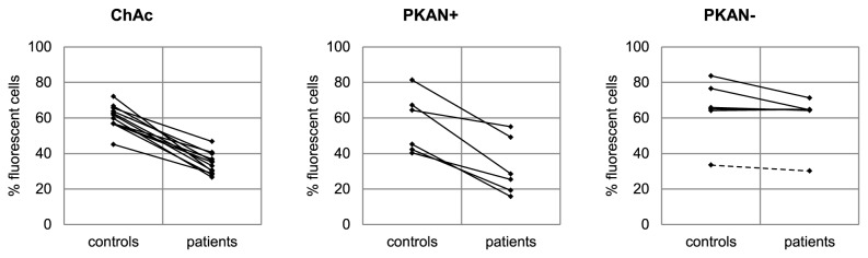
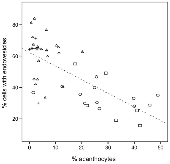
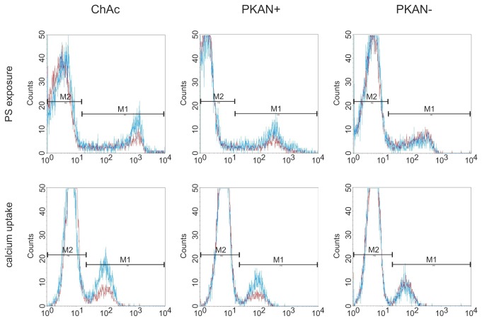
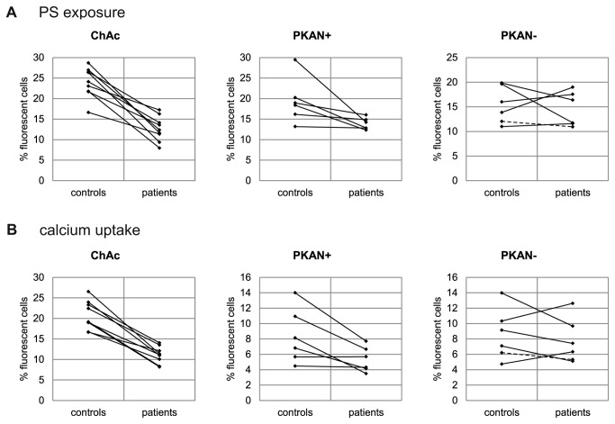
Neuroacanthocytosis (NA) refers to a group of heterogenous, rare genetic disorders, namely chorea acanthocytosis (ChAc), McLeod syndrome (MLS), Huntington's disease-like 2 (HDL2) and pantothenate kinase associated neurodegeneration (PKAN), that mainly affect the basal ganglia and are associated with similar neurological symptoms. PKAN is also assigned to a group of rare neurodegenerative diseases, known as NBIA (neurodegeneration with brain iron accumulation), associated with iron accumulation in the basal ganglia and progressive movement disorder. Acanthocytosis, the occurrence of misshaped erythrocytes with thorny protrusions, is frequently observed in ChAc and MLS patients but less prevalent in PKAN (about 10%) and HDL2 patients. The pathological factors that lead to the formation of the acanthocytic red blood cell shape are currently unknown. The aim of this study was to determine whether NA/NBIA acanthocytes differ in their functionality from normal erythrocytes. Several flow-cytometry-based assays were applied to test the physiological responses of the plasma membrane, namely drug-induced endocytosis, phosphatidylserine exposure and calcium uptake upon treatment with lysophosphatidic acid. ChAc red cell samples clearly showed a reduced response in drug-induced endovesiculation, lysophosphatidic acid-induced phosphatidylserine exposure, and calcium uptake. Impaired responses were also observed in acanthocyte-positive NBIA (PKAN) red cells but not in patient cells without shape abnormalities. These data suggest an "acanthocytic state" of the red cell where alterations in functional and interdependent membrane properties arise together with an acanthocytic cell shape. Further elucidation of the aberrant molecular mechanisms that cause this acanthocytic state may possibly help to evaluate the pathological pathways leading to neurodegeneration.
神经棘红细胞增多症(NA)是一组异质性的罕见遗传性疾病,包括舞蹈棘红细胞增多症(ChAc)、McLeod 综合征(MLS)、亨廷顿病样 2(HDL2)和泛酸激酶相关神经退行性变(PKAN),主要影响基底节,并伴有类似的神经症状。PKAN 也被归类为一组罕见的神经退行性疾病,称为 NBIA(脑铁蓄积相关神经变性),与基底节铁蓄积和进行性运动障碍有关。棘红细胞增多症,即畸形红细胞出现刺状突起,在 ChAc 和 MLS 患者中经常观察到,但在 PKAN(约 10%)和 HDL2 患者中较少见。导致棘状红细胞形成的病理因素目前尚不清楚。本研究旨在确定 NA/NBIA 棘红细胞在功能上是否与正常红细胞不同。应用几种基于流式细胞术的检测方法来测试细胞膜的生理反应,即药物诱导的内吞作用、磷脂酰丝氨酸暴露和溶血性磷脂酸处理后的钙摄取。ChAc 红细胞样本在药物诱导的内吞作用、溶血性磷脂酸诱导的磷脂酰丝氨酸暴露和钙摄取方面明显显示出反应降低。在棘红细胞阳性的 NBIA(PKAN)红细胞中也观察到受损的反应,但在没有形态异常的患者细胞中则没有。这些数据表明红细胞存在“棘状细胞状态”,其中功能和相互依赖的膜特性的改变与棘状细胞形态一起出现。进一步阐明导致这种棘状细胞状态的异常分子机制可能有助于评估导致神经退行性变的病理途径。