Halcsik Erik, Forni Maria Fernanda, Fujita Andre, Verano-Braga Thiago, Jensen Ole Nørregaard, Sogayar Mari Cleide
Chemistry Institute, Department of Biochemistry, Cell and Molecular Therapy Center (NUCEL/NETCEM), School of Medicine, University of São Paulo, São Paulo 05508-000, SP, Brazil.
BMC Cell Biol. 2013 Oct 22;14:47. doi: 10.1186/1471-2121-14-47.
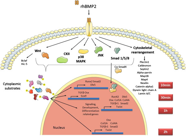
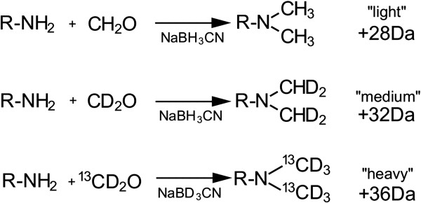
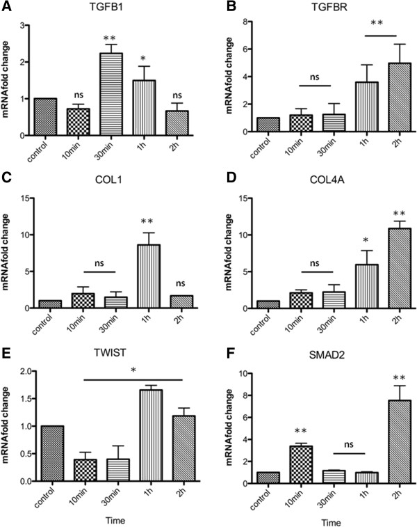
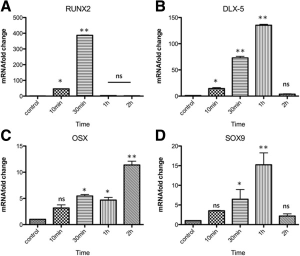
Bone fractures and loss represent significant costs for the public health system and often affect the patients quality of life, therefore, understanding the molecular basis for bone regeneration is essential. Cytokines, such as IL-6, IL-10 and TNFα, secreted by inflammatory cells at the lesion site, at the very beginning of the repair process, act as chemotactic factors for mesenchymal stem cells, which proliferate and differentiate into osteoblasts through the autocrine and paracrine action of bone morphogenetic proteins (BMPs), mainly BMP-2. Although it is known that BMP-2 binds to ActRI/BMPR and activates the SMAD 1/5/8 downstream effectors, little is known about the intracellular mechanisms participating in osteoblastic differentiation. We assessed differences in the phosphorylation status of different cellular proteins upon BMP-2 osteogenic induction of isolated murine skin mesenchymal stem cells using Triplex Stable Isotope Dimethyl Labeling coupled with LC/MS.
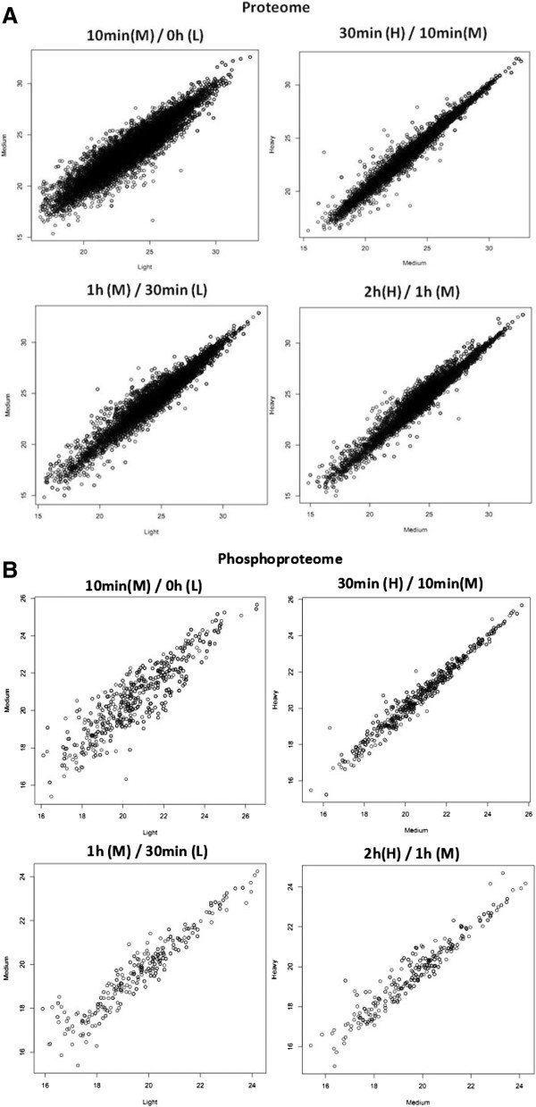
From 150 μg of starting material, 2,264 proteins were identified and quantified at five different time points, 235 of which are differentially phosphorylated. Kinase motif analysis showed that several substrates display phosphorylation sites for Casein Kinase, p38, CDK and JNK. Gene ontology analysis showed an increase in biological processes related with signaling and differentiation at early time points after BMP2 induction. Moreover, proteins involved in cytoskeleton rearrangement, Wnt and Ras pathways were found to be differentially phosphorylated during all timepoints studied.
Taken together, these data, allow new insights on the intracellular substrates which are phosphorylated early on during differentiation to BMP2-driven osteoblastic differentiation of skin-derived mesenchymal stem cells.
骨折和骨质流失给公共卫生系统带来了巨大成本,且常常影响患者的生活质量,因此,了解骨再生的分子基础至关重要。在修复过程开始时,损伤部位的炎症细胞分泌的细胞因子,如白细胞介素-6、白细胞介素-10和肿瘤坏死因子α,作为间充质干细胞的趋化因子,间充质干细胞通过骨形态发生蛋白(BMPs),主要是BMP-2的自分泌和旁分泌作用增殖并分化为成骨细胞。尽管已知BMP-2与激活素受体I型/BMP受体(ActRI/BMPR)结合并激活下游效应器SMAD 1/5/8,但对于参与成骨细胞分化的细胞内机制知之甚少。我们使用三重稳定同位素二甲基标记结合液相色谱/质谱法,评估了BMP-2对分离的小鼠皮肤间充质干细胞进行成骨诱导后不同细胞蛋白的磷酸化状态差异。
从150μg起始材料中,在五个不同时间点鉴定并定量了2264种蛋白质,其中235种蛋白质存在磷酸化差异。激酶基序分析表明,几种底物显示出酪蛋白激酶、p38、细胞周期蛋白依赖性激酶(CDK)和应激活化蛋白激酶(JNK)的磷酸化位点。基因本体分析表明,在BMP2诱导后的早期时间点,与信号传导和分化相关的生物学过程有所增加。此外,发现在所有研究时间点,参与细胞骨架重排、Wnt和Ras途径的蛋白质存在磷酸化差异。
综上所述,这些数据为皮肤来源的间充质干细胞向BMP2驱动成骨细胞分化过程中早期磷酸化的细胞内底物提供了新的见解。