Ackermann Maximilian, Houdek Jan P, Gibney Barry C, Ysasi Alexandra, Wagner Willi, Belle Janeil, Schittny Johannes C, Enzmann Frieder, Tsuda Akira, Mentzer Steven J, Konerding Moritz A
Institute of Functional and Clinical Anatomy, University Medical Center of the Johannes Gutenberg-University Mainz, 55128, Mainz, Germany.
Angiogenesis. 2014 Jul;17(3):541-51. doi: 10.1007/s10456-013-9399-9. Epub 2013 Oct 23.
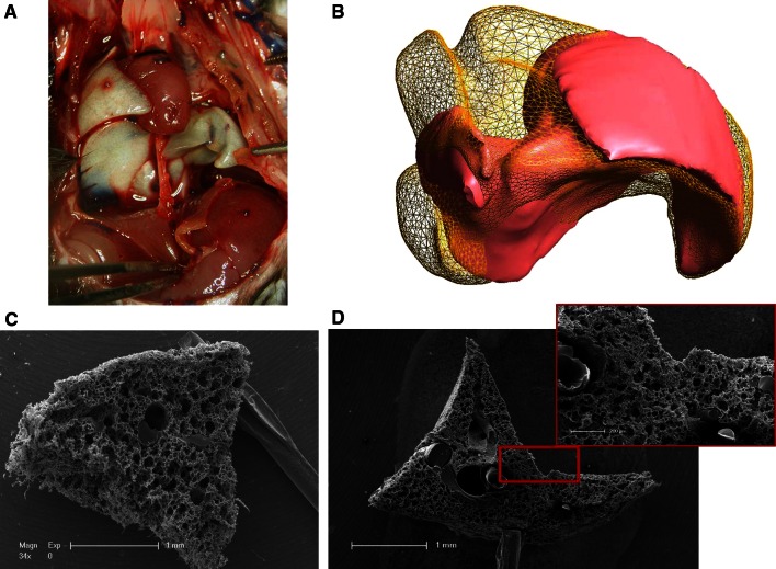
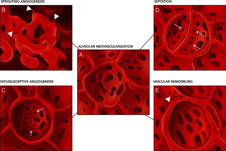
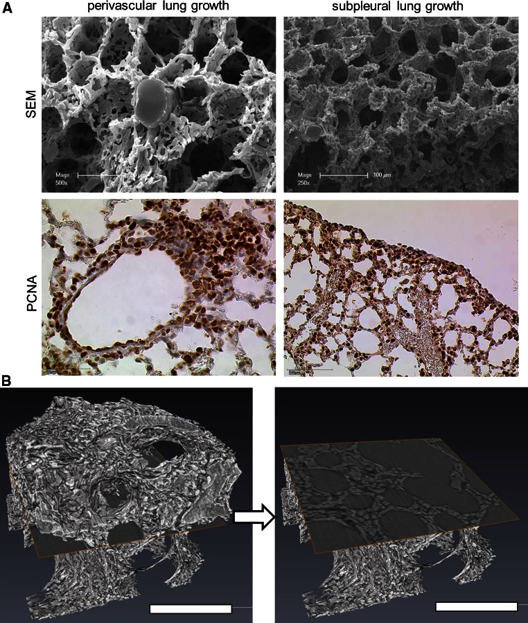
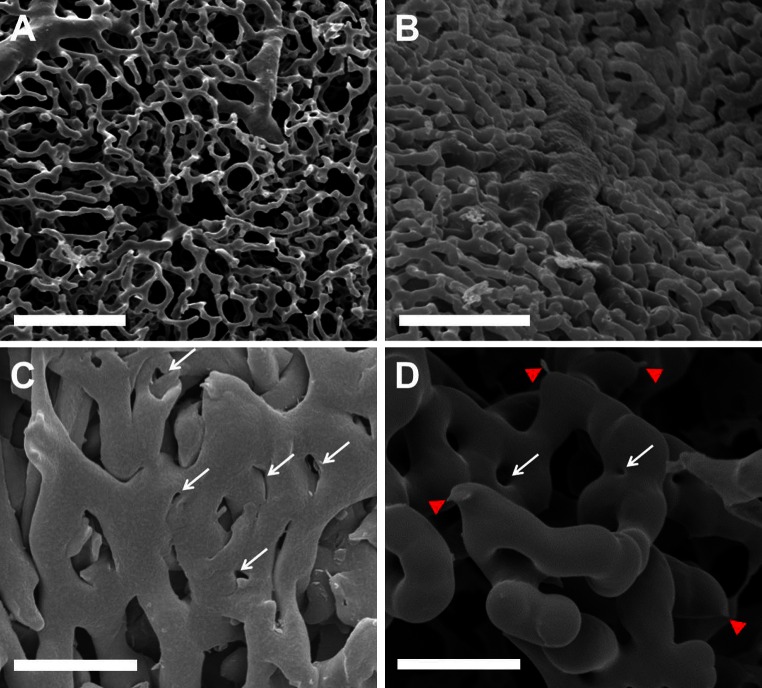
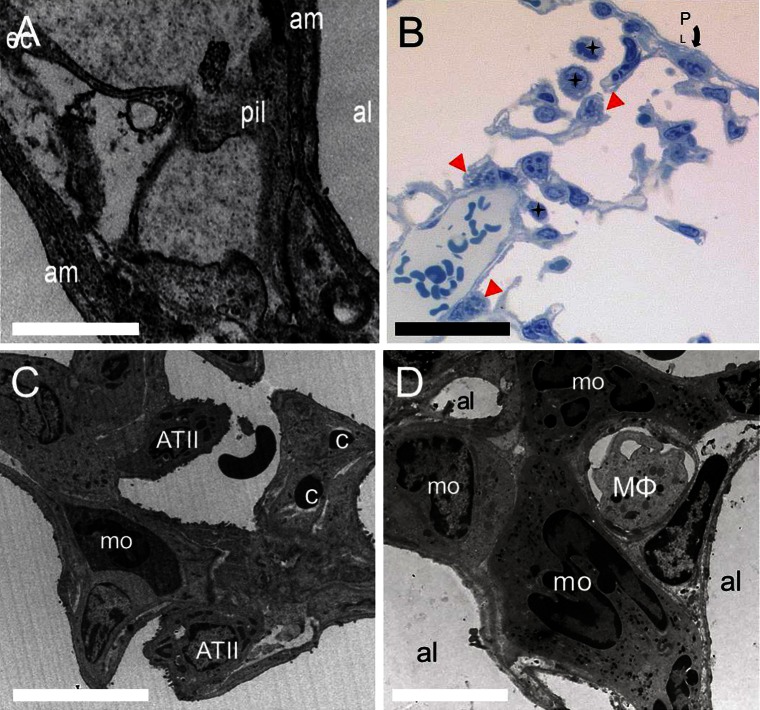
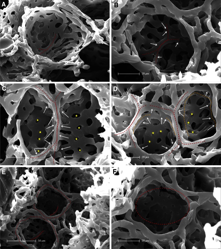
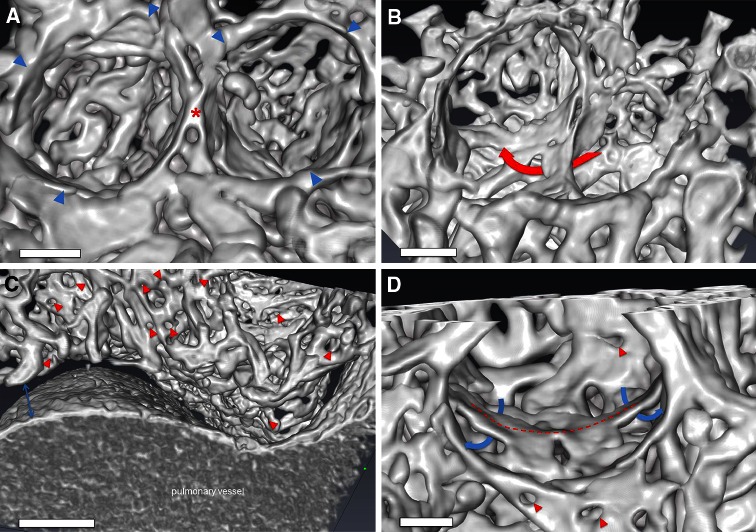
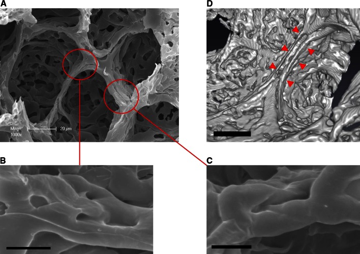
In most rodents and some other mammals, the removal of one lung results in compensatory growth associated with dramatic angiogenesis and complete restoration of lung capacity. One pivotal mechanism in neoalveolarization is neovascularization, because without angiogenesis new alveoli can not be formed. The aim of this study is to image and analyze three-dimensionally the different patterns of neovascularization seen following pneumonectomy in mice on a sub-micron-scale. C57/BL6 mice underwent a left-sided pneumonectomy. Lungs were harvested at various timepoints after pneumonectomy. Volume analysis by microCT revealed a striking increase of 143 percent in the cardiac lobe 14 days after pneumonectomy. Analysis of microvascular corrosion casting demonstrated spatially heterogenous vascular densitities which were in line with the perivascular and subpleural compensatory growth pattern observed in anti-PCNA-stained lung sections. Within these regions an expansion of the vascular plexus with increased pillar formations and sprouting angiogenesis, originating both from pre-existing bronchial and pulmonary vessels was observed. Also, type II pneumocytes and alveolar macrophages were seen to participate actively in alveolar neo-angiogenesis after pneumonectomy. 3D-visualizations obtained by high-resolution synchrotron radiation X-ray tomographic microscopy showed the appearance of double-layered vessels and bud-like alveolar baskets as have already been described in normal lung development. Scanning electron microscopy data of microvascular architecture also revealed a replication of perialveolar vessel networks through septum formation as already seen in developmental alveolarization. In addition, the appearance of pillar formations and duplications on alveolar entrance ring vessels in mature alveoli are indicative of vascular remodeling. These findings indicate that sprouting and intussusceptive angiogenesis are pivotal mechanisms in adult lung alveolarization after pneumonectomy. Various forms of developmental neoalveolarization may also be considered to contribute in compensatory lung regeneration.
在大多数啮齿动物和其他一些哺乳动物中,切除一侧肺会导致代偿性生长,伴有显著的血管生成,并使肺容量完全恢复。新肺泡形成的一个关键机制是新血管形成,因为没有血管生成就无法形成新的肺泡。本研究的目的是在亚微米尺度上对小鼠肺切除术后出现的不同新血管形成模式进行三维成像和分析。C57/BL6小鼠接受了左侧肺切除术。在肺切除术后的不同时间点采集肺组织。通过微型计算机断层扫描(microCT)进行的体积分析显示,肺切除术后14天,心叶体积显著增加了143%。微血管铸型分析表明,血管密度在空间上存在异质性,这与抗增殖细胞核抗原(anti-PCNA)染色的肺切片中观察到的血管周围和胸膜下代偿性生长模式一致。在这些区域内,观察到血管丛扩张,柱状结构增加,出现了源自既存支气管血管和肺血管的芽生血管生成。此外,还观察到II型肺泡上皮细胞和肺泡巨噬细胞在肺切除术后积极参与肺泡新生血管生成。通过高分辨率同步辐射X射线断层显微镜获得的三维可视化图像显示了双层血管和芽状肺泡篮的出现,这在正常肺发育中已有描述。微血管结构的扫描电子显微镜数据也显示,通过隔膜形成复制了肺泡周围血管网络,这在发育性肺泡化中也已见到。此外,成熟肺泡中肺泡入口环血管上柱状结构和重复结构的出现表明存在血管重塑。这些发现表明,芽生血管生成和套叠式血管生成是肺切除术后成年肺肺泡化的关键机制。各种形式的发育性新肺泡形成也可能被认为有助于肺的代偿性再生。