Department of Medical Oncology, VU University Medical Center, De Boelelaan 1117, 1081HV, Amsterdam, Netherlands.
Department of Pharmacy, University of Pisa, Pisa, Italy.
Br J Cancer. 2014 Jan 7;110(1):172-82. doi: 10.1038/bjc.2013.681. Epub 2013 Oct 31.
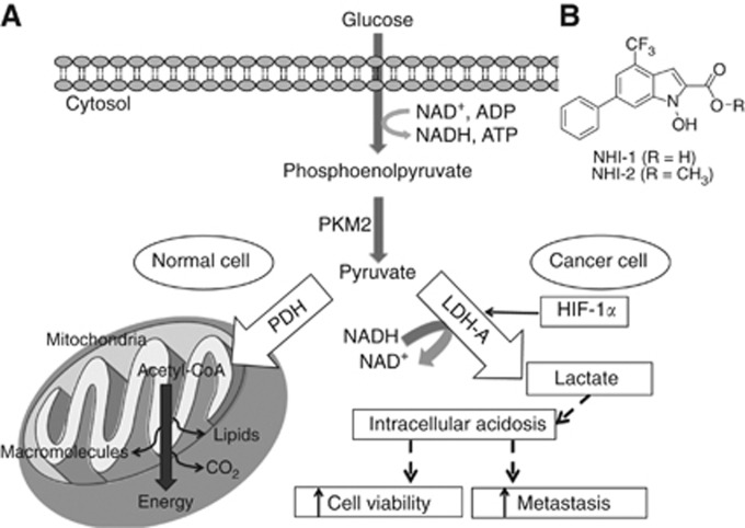
Hypoxia is a driving force in pancreatic-ductal-adenocarcinoma (PDAC) growth, metastasis and chemoresistance. The muscle-isoform of lactate dehydrogenase (LDH-A) constitutes a major checkpoint for the switch to anaerobic glycolysis, ensuring supply of energy and anabolites in hypoxic-environments. Therefore, we investigated the molecular mechanisms underlying the pharmacological interaction of novel LDH-A inhibitors in combination with gemcitabine in PDAC cells.
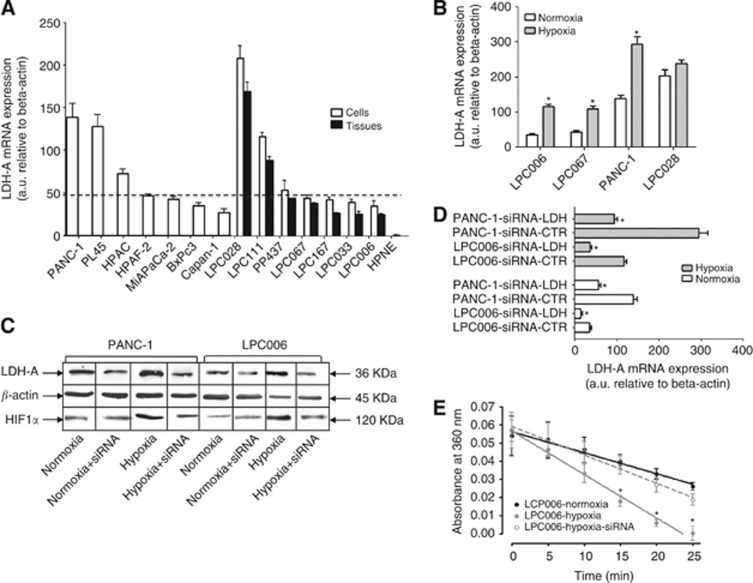
Lactate dehydrogenase A levels were studied by quantitative RT-PCR, western blot, immunofluorescence and activity assays in 14 PDAC cells, including primary-cell-cultures and spheroids, in normoxic and hypoxic conditions. Cell proliferation, migration and key determinants of drug activity were evaluated by sulforhodamine-B-assay, wound-healing assay, PCR and LC-MS/MS.
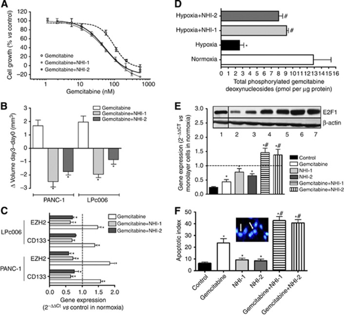
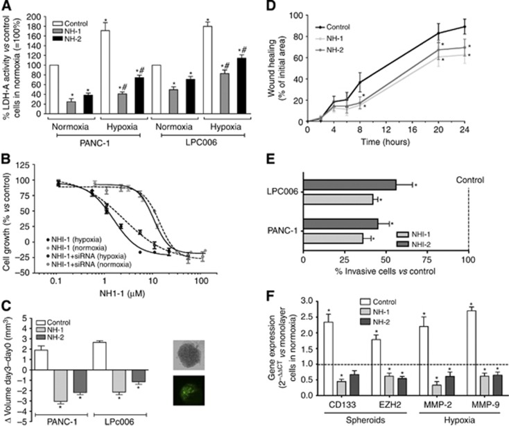
Lactate dehydrogenase A was significantly increased under hypoxic conditions (1% O2), where the novel LDH-A inhibitors proved to be particularly effective (e.g., with IC50 values of 0.9 vs 16.3 μM for NHI-1 in LPC006 in hypoxia vs normoxia, respectively). These compounds induced apoptosis, affected invasiveness and spheroid-growth, reducing expression of metalloproteinases and cancer-stem-like-cells markers (CD133+). Their synergistic interaction with gemcitabine, with combination index values <0.4 in hypoxia, might also be attributed to modulation of gemcitabine metabolism, overcoming the reduced synthesis of phosphorylated metabolites.
Lactate dehydrogenase A is a viable target in PDAC, and novel LDH-A inhibitors display synergistic cytotoxic activity with gemcitabine, offering an innovative tool in hypoxic tumours.
缺氧是胰腺导管腺癌(PDAC)生长、转移和化疗耐药的驱动力。肌肉同工型乳酸脱氢酶(LDH-A)构成了向无氧糖酵解转变的主要检查点,确保在缺氧环境中供应能量和代谢物。因此,我们研究了新型 LDH-A 抑制剂与吉西他滨联合在 PDAC 细胞中相互作用的分子机制。
在常氧和缺氧条件下,通过定量 RT-PCR、western blot、免疫荧光和活性测定,在 14 种 PDAC 细胞(包括原代细胞培养物和球体)中研究 LDH-A 水平。通过磺酰罗丹明 B 测定法、划痕愈合测定法、PCR 和 LC-MS/MS 评估细胞增殖、迁移和药物活性的关键决定因素。
在缺氧条件下(1% O2),LDH-A 显著增加,新型 LDH-A 抑制剂在此条件下特别有效(例如,在缺氧条件下,NHI-1 的 IC50 值为 0.9 μM,而 LPC006 为 16.3 μM)。这些化合物诱导细胞凋亡,影响侵袭性和球体生长,降低金属蛋白酶和癌症干细胞样细胞标志物(CD133+)的表达。它们与吉西他滨的协同相互作用,在缺氧条件下的组合指数值<0.4,也可能归因于调节吉西他滨代谢,克服磷酸化代谢物合成减少。
LDH-A 是 PDAC 的一个可行靶点,新型 LDH-A 抑制剂与吉西他滨具有协同细胞毒性作用,为缺氧肿瘤提供了一种创新工具。