Massihnia Daniela, Avan Amir, Funel Niccola, Maftouh Mina, van Krieken Anne, Granchi Carlotta, Raktoe Rajiv, Boggi Ugo, Aicher Babette, Minutolo Filippo, Russo Antonio, Leon Leticia G, Peters Godefridus J, Giovannetti Elisa
Department of Medical Oncology VU University Medical Center, Cancer Center Amsterdam, CCA room 1.52, De Boelelaan 1117, 1081 HV, Amsterdam, The Netherlands.
Department of Surgical, Oncological and Oral Sciences, Section of Medical Oncology, University of Palermo, Palermo, Italy.
J Hematol Oncol. 2017 Jan 6;10(1):9. doi: 10.1186/s13045-016-0371-1.
There is increasing evidence of a constitutive activation of Akt in pancreatic ductal adenocarcinoma (PDAC), associated with poor prognosis and chemoresistance. Therefore, we evaluated the expression of phospho-Akt in PDAC tissues and cells, and investigated molecular mechanisms influencing the therapeutic potential of Akt inhibition in combination with gemcitabine.
Phospho-Akt expression was evaluated by immunohistochemistry in tissue microarrays (TMAs) with specimens tissue from radically-resected patients (n = 100). Data were analyzed by Fisher and log-rank test. In vitro studies were performed in 14 PDAC cells, including seven primary cultures, characterized for their Akt1 mRNA and phospho-Akt/Akt levels by quantitative-RT-PCR and immunocytochemistry. Growth inhibitory effects of Akt inhibitors and gemcitabine were evaluated by SRB assay, whereas modulation of Akt and phospho-Akt was investigated by Western blotting and ELISA. Cell cycle perturbation, apoptosis-induction, and anti-migratory behaviors were studied by flow cytometry, AnnexinV, membrane potential, and migration assay, while pharmacological interaction with gemcitabine was determined with combination index (CI) method.
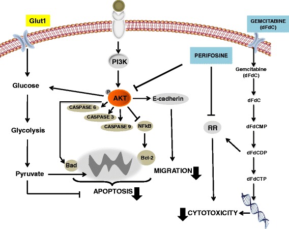
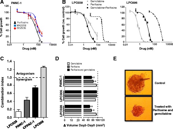
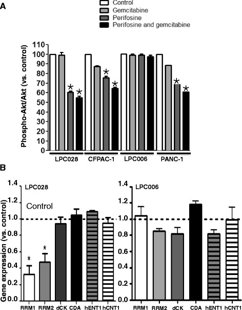
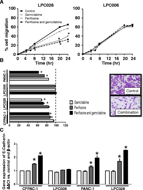
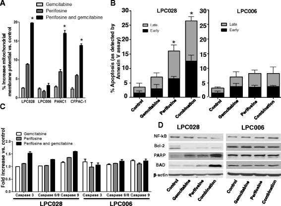
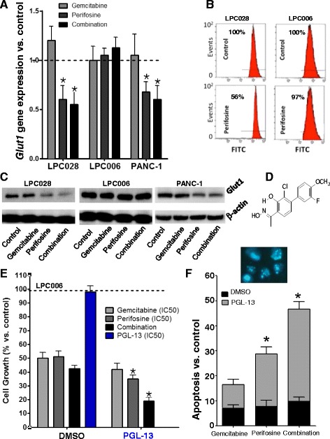
Immunohistochemistry of TMAs revealed a correlation between phospho-Akt expression and worse outcome, particularly in patients with the highest phospho-Akt levels, who had significantly shorter overall and progression-free-survival. Similar expression levels were detected in LPC028 primary cells, while LPC006 were characterized by low phospho-Akt. Remarkably, Akt inhibitors reduced cancer cell growth in monolayers and spheroids and synergistically enhanced the antiproliferative activity of gemcitabine in LPC028, while this combination was antagonistic in LPC006 cells. The synergistic effect was paralleled by a reduced expression of ribonucleotide reductase, potentially facilitating gemcitabine cytotoxicity. Inhibition of Akt decreased cell migration and invasion, which was additionally reduced by the combination with gemcitabine. This combination significantly increased apoptosis, associated with induction of caspase-3/6/8/9, PARP and BAD, and inhibition of Bcl-2 and NF-kB in LPC028, but not in LPC006 cells. However, targeting the key glucose transporter Glut1 resulted in similar apoptosis induction in LPC006 cells.
These data support the analysis of phospho-Akt expression as both a prognostic and a predictive biomarker, for the rational development of new combination therapies targeting the Akt pathway in PDAC. Finally, inhibition of Glut1 might overcome resistance to these therapies and warrants further studies.
越来越多的证据表明,Akt在胰腺导管腺癌(PDAC)中存在组成性激活,这与预后不良和化疗耐药相关。因此,我们评估了磷酸化Akt在PDAC组织和细胞中的表达,并研究了影响Akt抑制与吉西他滨联合治疗潜力的分子机制。
通过免疫组织化学对来自根治性切除患者的组织芯片(TMAs)(n = 100)中的标本组织进行磷酸化Akt表达评估。数据采用Fisher检验和对数秩检验进行分析。在14种PDAC细胞中进行体外研究,包括7种原代培养细胞,通过定量逆转录PCR和免疫细胞化学对其Akt1 mRNA以及磷酸化Akt/Akt水平进行表征。通过SRB法评估Akt抑制剂和吉西他滨的生长抑制作用,而通过蛋白质免疫印迹法和酶联免疫吸附测定法研究Akt和磷酸化Akt的调节情况。通过流式细胞术、膜联蛋白V、膜电位和迁移试验研究细胞周期扰动、凋亡诱导和抗迁移行为,同时用联合指数(CI)法确定与吉西他滨的药理相互作用。
TMAs的免疫组织化学显示磷酸化Akt表达与较差的预后相关,特别是在磷酸化Akt水平最高的患者中,这些患者的总生存期和无进展生存期明显较短。在LPC028原代细胞中检测到类似的表达水平,而LPC006细胞的特征是磷酸化Akt水平较低。值得注意的是,Akt抑制剂可降低单层和球体中的癌细胞生长,并协同增强吉西他滨在LPC028中的抗增殖活性,而这种联合在LPC006细胞中具有拮抗作用。这种协同作用伴随着核糖核苷酸还原酶表达的降低,可能促进了吉西他滨的细胞毒性。抑制Akt可降低细胞迁移和侵袭,与吉西他滨联合使用时这种作用进一步降低。这种联合显著增加了凋亡,与LPC028中半胱天冬酶-3/6/8/9、聚(ADP-核糖)聚合酶(PARP)和BAD的诱导以及Bcl-2和核因子κB的抑制相关,但在LPC006细胞中未出现这种情况。然而,靶向关键葡萄糖转运蛋白Glut1在LPC006细胞中导致了类似的凋亡诱导。
这些数据支持将磷酸化Akt表达分析作为一种预后和预测生物标志物,用于合理开发针对PDAC中Akt通路的新联合疗法。最后,抑制Glut1可能克服对这些疗法的耐药性,值得进一步研究。