Multimodal Imaging Physics Group, University of Applied Sciences Koblenz, RheinAhrCampus Remagen, 53424 Remagen, Germany ; Institute for Medical Engineering and Information Processing, MTI Mittelrhein, University of Koblenz, 56070 Koblenz, Germany.
Neuroimage Clin. 2012 Oct 5;1(1):121-30. doi: 10.1016/j.nicl.2012.09.013. eCollection 2012.
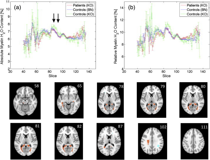
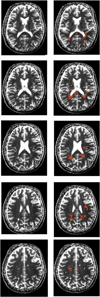
The current study investigates the whole brain myelin water content distribution applying a new approach that allows for the simultaneous mapping of total and relative myelin water content, T 1 and T 2* with full brain coverage and high resolution (1 × 1 × 2 mm(3)). The data was collected at two different sites in healthy controls to validate the independence of a specific setup. In addition, a group of patients with known white matter affections was investigated to compare two measures of myelin, i.e. relative and absolute myelin water content. Based on the first dataset, a quantitative myelin water content atlas was created which served as a control set for the other two datasets. Both control groups measured at different institutions yielded consistent results. However, distinct regions of reduced myelin water content were observed for the patient dataset, both on an individual basis and in a group-wise comparison. The comparison between the absolute and relative measurement of myelin water content in MS patients showed that the relative measurement, which is employed by many researchers, overestimates both disease volume and the corresponding reduction of myelin water content in white matter lesions. However, for normal appearing white matter, no difference between both approaches was detected. The results obtained in the current study demonstrate that absolute myelin water content can reliably be determined in a multicentre environment using standard MR sequences. The optimised protocol allows for a measurement of four quantitative parameters with full brain coverage in only 10 min. This might expedite a more widespread future use of quantitative MRI methods for clinical research and diagnosis.
本研究应用一种新方法,研究全脑髓鞘水含量分布,该方法允许同时绘制总髓鞘水含量和相对髓鞘水含量、T 1 和 T 2*,覆盖整个大脑,具有高分辨率(1×1×2mm 3)。该数据是在两个不同的健康对照组地点收集的,以验证特定设置的独立性。此外,还对一组已知的白质病变患者进行了研究,比较了两种髓鞘测量方法,即相对和绝对髓鞘水含量。基于第一个数据集,创建了一个定量髓鞘水含量图谱,作为另外两个数据集的对照组。在不同机构测量的两个对照组均产生了一致的结果。然而,对于患者数据集,在个体和组间比较中,都观察到明显的髓鞘水含量降低区域。MS 患者的髓鞘水含量绝对和相对测量的比较表明,相对测量(许多研究人员采用的方法)高估了疾病体积以及白质病变中髓鞘水含量的相应减少。然而,对于正常表现的白质,两种方法之间没有差异。本研究的结果表明,绝对髓鞘水含量可以在多中心环境中使用标准磁共振序列可靠地确定。优化的方案允许在仅 10 分钟内对全脑覆盖的四个定量参数进行测量。这可能会加速定量 MRI 方法在临床研究和诊断中的更广泛应用。