Department of Medicine and Biomedical Engineering, Translational Imaging in Neurology Basel, University Hospital Basel and University of Basel, Basel, Switzerland.
Departments of Medicine, Clinical Research and Biomedical Engineering Neurologic Clinic and Policlinic, Switzerland, University Hospital Basel and University of Basel, Basel, Switzerland.
Brain. 2021 Jul 28;144(6):1684-1696. doi: 10.1093/brain/awab088.
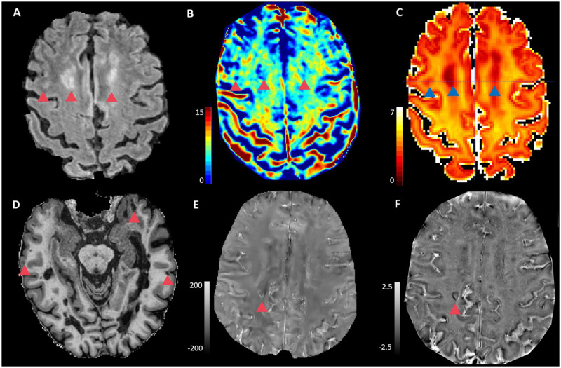
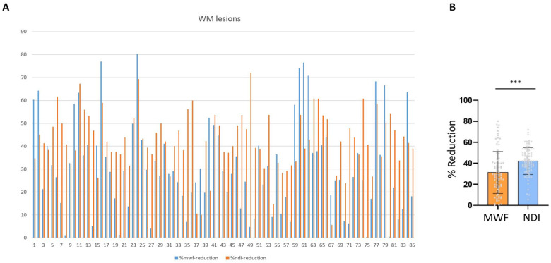
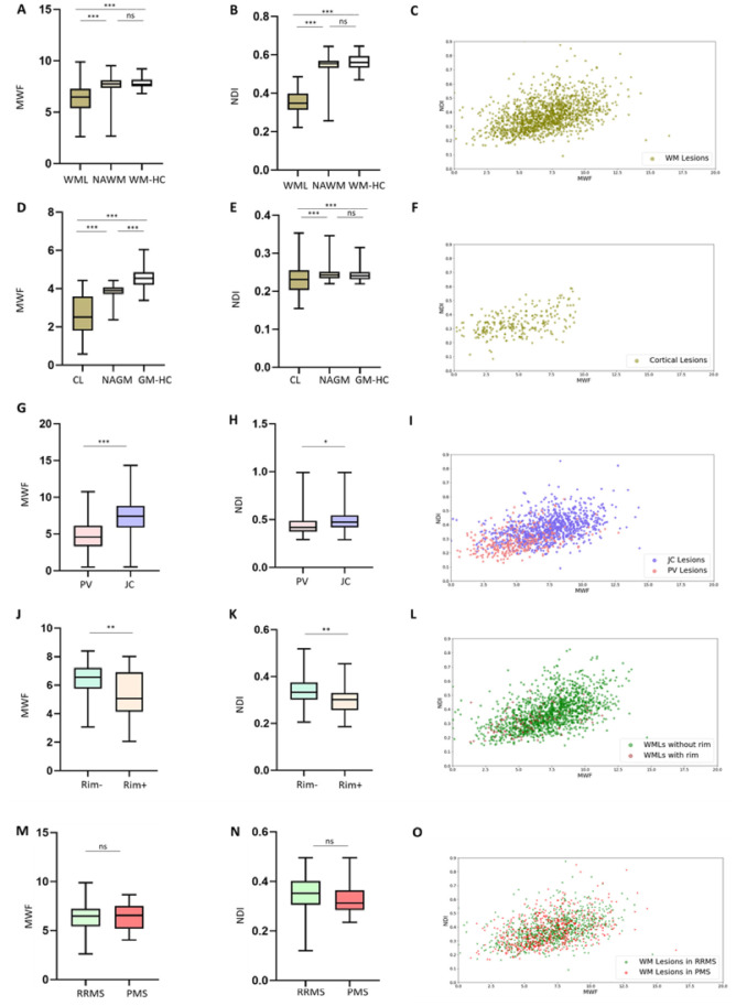
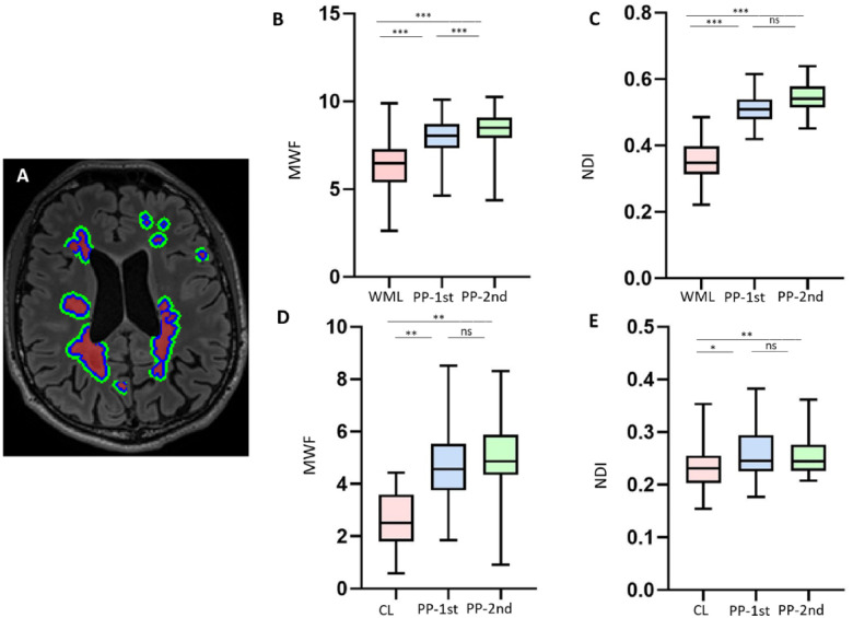
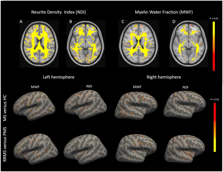
Damage to the myelin sheath and the neuroaxonal unit is a cardinal feature of multiple sclerosis; however, a detailed characterization of the interaction between myelin and axon damage in vivo remains challenging. We applied myelin water and multi-shell diffusion imaging to quantify the relative damage to myelin and axons (i) among different lesion types; (ii) in normal-appearing tissue; and (iii) across multiple sclerosis clinical subtypes and healthy controls. We also assessed the relation of focal myelin/axon damage with disability and serum neurofilament light chain as a global biological measure of neuroaxonal damage. Ninety-one multiple sclerosis patients (62 relapsing-remitting, 29 progressive) and 72 healthy controls were enrolled in the study. Differences in myelin water fraction and neurite density index were substantial when lesions were compared to healthy control subjects and normal-appearing multiple sclerosis tissue: both white matter and cortical lesions exhibited a decreased myelin water fraction and neurite density index compared with healthy (P < 0.0001) and peri-plaque white matter (P < 0.0001). Periventricular lesions showed decreased myelin water fraction and neurite density index compared with lesions in the juxtacortical region (P < 0.0001 and P < 0.05). Similarly, lesions with paramagnetic rims showed decreased myelin water fraction and neurite density index relative to lesions without a rim (P < 0.0001). Also, in 75% of white matter lesions, the reduction in neurite density index was higher than the reduction in the myelin water fraction. Besides, normal-appearing white and grey matter revealed diffuse reduction of myelin water fraction and neurite density index in multiple sclerosis compared to healthy controls (P < 0.01). Further, a more extensive reduction in myelin water fraction and neurite density index in normal-appearing cortex was observed in progressive versus relapsing-remitting participants. Neurite density index in white matter lesions correlated with disability in patients with clinical deficits (P < 0.01, beta = -10.00); and neurite density index and myelin water fraction in white matter lesions were associated to serum neurofilament light chain in the entire patient cohort (P < 0.01, beta = -3.60 and P < 0.01, beta = 0.13, respectively). These findings suggest that (i) myelin and axon pathology in multiple sclerosis is extensive in both lesions and normal-appearing tissue; (ii) particular types of lesions exhibit more damage to myelin and axons than others; (iii) progressive patients differ from relapsing-remitting patients because of more extensive axon/myelin damage in the cortex; and (iv) myelin and axon pathology in lesions is related to disability in patients with clinical deficits and global measures of neuroaxonal damage.
髓鞘和神经轴突单位的损伤是多发性硬化症的主要特征;然而,在体内详细描述髓鞘和轴突损伤之间的相互作用仍然具有挑战性。我们应用髓鞘水和多壳扩散成像来定量评估不同病变类型之间、正常表现组织之间以及多发性硬化症的不同临床亚型和健康对照组之间髓鞘和轴突的相对损伤。我们还评估了局灶性髓鞘/轴突损伤与残疾之间的关系,以及血清神经丝轻链作为神经轴突损伤的整体生物标志物。91 名多发性硬化症患者(62 名复发缓解型,29 名进展型)和 72 名健康对照组参与了这项研究。与健康对照组和正常表现的多发性硬化组织相比,病变之间的髓鞘水分数和神经丝密度指数差异显著:白质和皮质病变的髓鞘水分数和神经丝密度指数均低于健康对照组(P < 0.0001)和斑块周围白质(P < 0.0001)。脑室周围病变的髓鞘水分数和神经丝密度指数低于皮质旁病变(P < 0.0001 和 P < 0.05)。同样,有顺磁性边缘的病变与无边缘的病变相比,髓鞘水分数和神经丝密度指数均降低(P < 0.0001)。此外,在 75%的白质病变中,神经丝密度指数的降低高于髓鞘水分数的降低。此外,与健康对照组相比,正常表现的白质和灰质中髓鞘水分数和神经丝密度指数均有弥漫性降低(P < 0.01)。此外,与复发缓解型患者相比,进展型患者的正常表现皮层中的髓鞘水分数和神经丝密度指数下降更为明显。有临床缺陷的患者的白质病变中的神经丝密度指数与残疾相关(P < 0.01,β = -10.00);白质病变中的神经丝密度指数和髓鞘水分数与整个患者队列的血清神经丝轻链相关(P < 0.01,β = -3.60 和 P < 0.01,β = 0.13)。这些发现表明:(i)多发性硬化症中的髓鞘和轴突病理学在病变和正常表现组织中都很广泛;(ii)特定类型的病变比其他病变对髓鞘和轴突的损伤更大;(iii)进展型患者与复发缓解型患者不同,因为皮层中轴突/髓鞘损伤更为广泛;(iv)病变中的髓鞘和轴突病理学与有临床缺陷的患者的残疾以及神经轴突损伤的整体生物标志物相关。