Arliani Gustavo Gonçalves, Astur Diego Costa, Moraes Eduardo Ramalho, Kaleka Camila Cohen, Jalikjian Wahi, Golano Pau, Cohen Moisés
Centro de Traumatologia do Esporte (CETE), Departamento de Ortopedia e Traumatologia da Universidade Federal de São Paulo SP, Brazil (DOT-UNIFESP/EPM).
Open Access J Sports Med. 2012 Nov 12;3:183-8. doi: 10.2147/OAJSM.S37203. eCollection 2012.
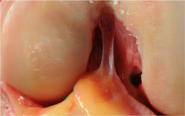
The anterior cruciate ligament (ACL) is an important structure in the knee. The ACL does not heal following lesions, and surgical reconstruction is the standard treatment among athletes. Some steps of ACL reconstruction remain controversial. It is important to fully understand the anatomy of the ACL to accurately reproduce its anatomy during surgical reconstructions. The purpose of this study was to evaluate the use of anaglyphic images that produce 3D images to better visualize the anatomy of the ACL, and to highlight the anatomical features of this ligament as reported in the literature.
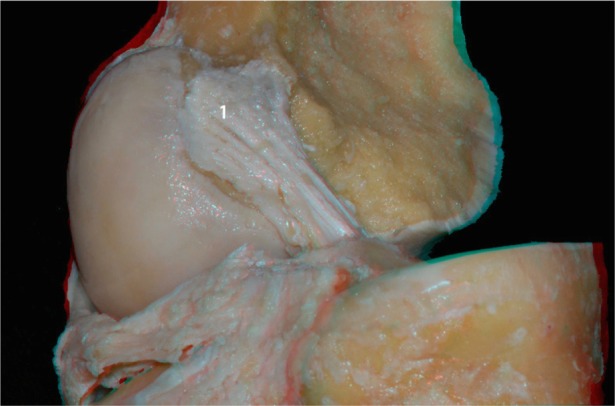
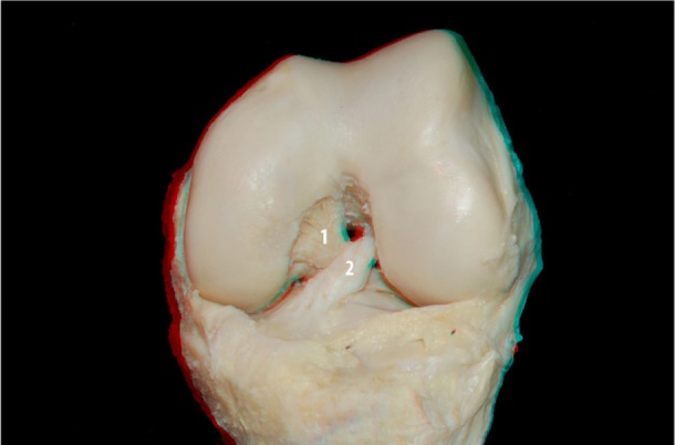
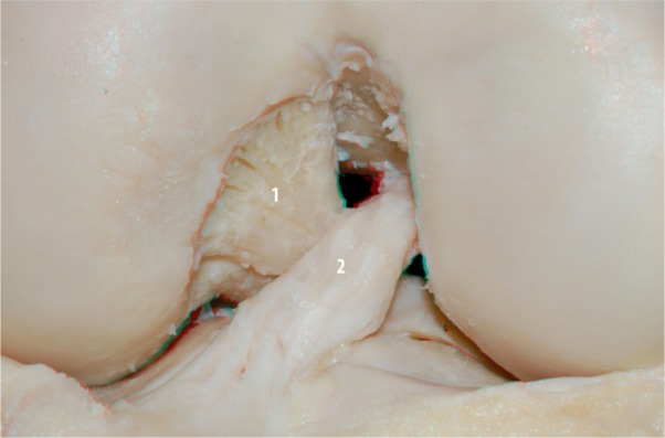
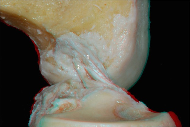
We included ten knees in this study. After dissection of the knee structures, pictures were acquired using a camera with Nikon D40, AF-S Nikkor 18-55 mm (1:3.5-5.6 G2 ED), and Micro Nikkor 105 mm (1:2.8) lenses. The pair of images was processed using Callipyan 3D Anabuilder software, which transforms the two images into one anaglyphic image.
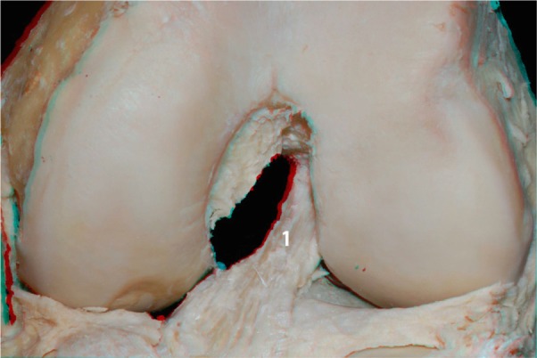
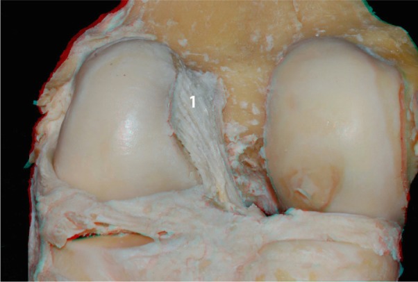
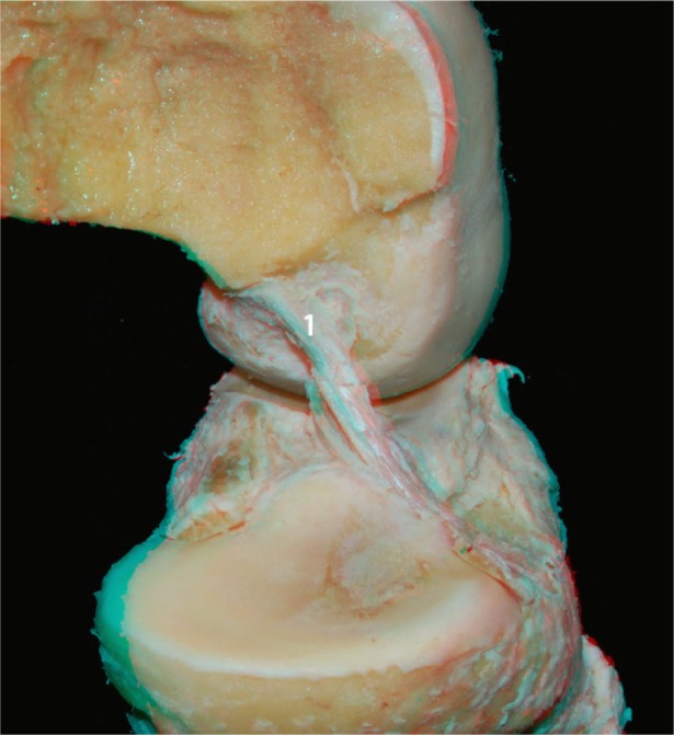
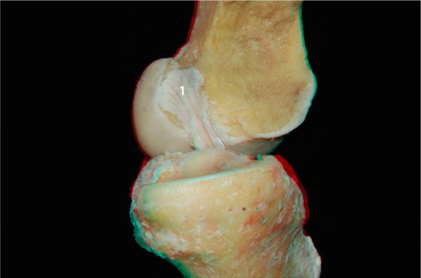
During the dissection of the knees, nine pictures were acquired and transformed into anaglyphic images.
This study, demonstrated that the use of 3D images is a useful tool that can improve the knowledge of the anatomy of the knee, while also facilitating knee reconstruction surgery.
前交叉韧带(ACL)是膝关节的重要结构。ACL损伤后无法自行愈合,手术重建是运动员的标准治疗方法。ACL重建的一些步骤仍存在争议。在手术重建过程中,充分了解ACL的解剖结构以准确重现其解剖结构非常重要。本研究的目的是评估使用产生3D图像的互补色立体图像以更好地可视化ACL的解剖结构,并突出文献中报道的该韧带的解剖特征。
本研究纳入了10个膝关节。在解剖膝关节结构后,使用配备尼康D40相机、AF-S尼克尔18 - 55毫米(1:3.5 - 5.6 G2 ED)镜头和Micro尼克尔105毫米(1:2.8)镜头的相机采集图片。使用Callipyan 3D Anabuilder软件处理这对图像,该软件将两幅图像转换为一幅互补色立体图像。
在膝关节解剖过程中,采集了9张图片并转换为互补色立体图像。
本研究表明,使用3D图像是一种有用的工具,它可以增进对膝关节解剖结构的了解,同时也有助于膝关节重建手术。