Department of Restorative Dentistry, São Leopoldo Mandic Institute and Research Center, CampinasSP, Brazil.
J Appl Oral Sci. 2013 Sep-Oct;21(5):452-9. doi: 10.1590/1679-775720130120.
The aim of this in vitro study was to evaluate the microtensile bond strength (µTBS) to dentin of two different restorative systems: silorane-based (P90), and methacrylate-based (P60), using two cavity models.
Occlusal enamel of 40 human third molars was removed to expose flat dentin surface. Class I cavities with 4 mm mesial-distal width, 3 mm buccal-lingual width and 3 mm depth (C-factor=4.5) were prepared in 20 teeth, which were divided into two groups (n=10) restored with P60 and P90, bulk-filled after dentin treatment according to manufacturer's instructions. Flat buccal dentin surfaces were prepared in the 20 remaining teeth (C-factor=0.2) and restored with resin blocks measuring 4x3x3 mm using the two restorative systems (n=10). The teeth were sectioned into samples with area between 0.85 and 1.25 mm2 that were submitted to µTBS testing, using a universal testing machine (EMIC) at speed of 0.5 mm/min. Fractured specimens were analyzed under stereomicroscope and categorized according to fracture pattern. Data were analyzed using ANOVA and Tukey Kramer tests.
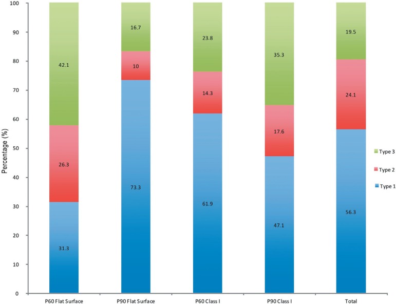
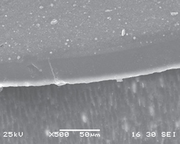
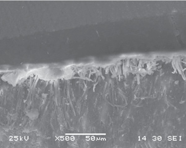
For flat surfaces, P60 obtained higher bond strength values compared with P90. However, for Class I cavities, P60 showed significant reduction in bond strength (p<0.05). No statistical difference between restorative systems was shown for Class I cavity model (p>0.05), or between Class I Cavity and Flat Surface group, considering P90 restorative system (p>0.05). Regarding fracture pattern, there was no statistical difference among groups (p=0.0713) and 56.3% of the fractures were adhesive.
It was concluded that methacrylate-based composite µTBS was influenced by cavity models, and the use of silorane-based composite led to similar bond strength values compared to the methacrylate-based composite in cavities with high C-factor.
本体外研究旨在评估两种不同修复系统(硅烷基 P90 和甲基丙烯酸酯基 P60)对牙本质的微拉伸粘结强度(µTBS),使用两种洞型模型。
从 40 个人类第三磨牙中去除咬合面釉质,以暴露平坦的牙本质表面。在 20 颗牙齿中制备 4mm 近远中宽度、3mm 颊舌宽度和 3mm 深度的 I 类洞(C 因子=4.5),根据制造商的说明,在牙本质处理后,将这些牙齿分为 P60 和 P90 两组进行大块填充。在剩余的 20 颗牙齿中制备平坦的颊侧牙本质表面(C 因子=0.2),并用两种修复系统(每组 n=10)填充 4x3x3mm 的树脂块。将牙齿切成面积为 0.85 至 1.25mm2 的样本,使用万能试验机(EMIC)以 0.5mm/min 的速度进行 µTBS 测试。在立体显微镜下分析断裂样本,并根据断裂模式进行分类。使用方差分析和 Tukey Kramer 检验对数据进行分析。
对于平坦表面,P60 获得的粘结强度值高于 P90。然而,对于 I 类洞,P60 的粘结强度显著降低(p<0.05)。对于 I 类洞模型(p>0.05)或 P90 修复系统的平坦表面组(p>0.05),两种修复系统之间没有显示出统计学差异。关于断裂模式,各组之间没有统计学差异(p=0.0713),56.3%的断裂为黏附性断裂。
甲基丙烯酸酯基复合材料的 µTBS 受洞型模型的影响,而使用硅烷基复合材料在 C 因子较高的腔中导致与甲基丙烯酸酯基复合材料相似的粘结强度值。