Liu Xiao-Wen, Cai Tian-Yu, Zhu Hong, Cao Ji, Su Yi, Hu Yong-Zhou, He Qiao-Jun, Yang Bo
Zhejiang Province Key Laboratory of Anti-Cancer Drug Research; Institute of Pharmacology and Toxicology; College of Pharmaceutical Sciences; Zhejiang University; Hangzhou, China.
ZJU-ENS Joint laboratory of Medicinal Chemistry; College of Pharmaceutical Sciences; Zhejiang University; Hangzhou, China.
Autophagy. 2014 Jan;10(1):111-22. doi: 10.4161/auto.26838. Epub 2013 Nov 11.
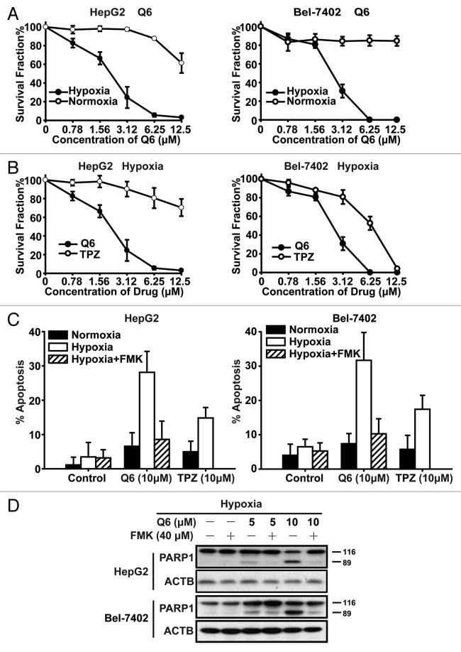
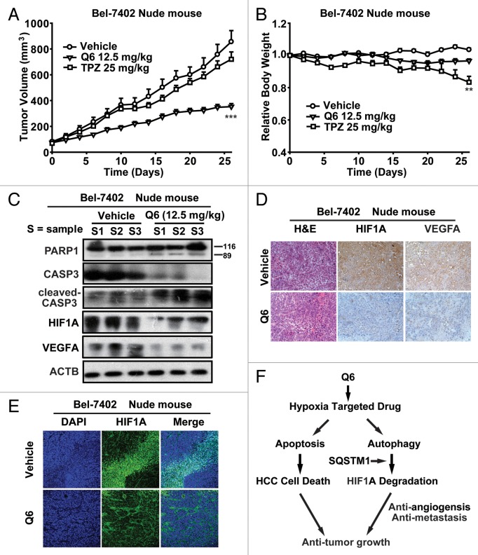
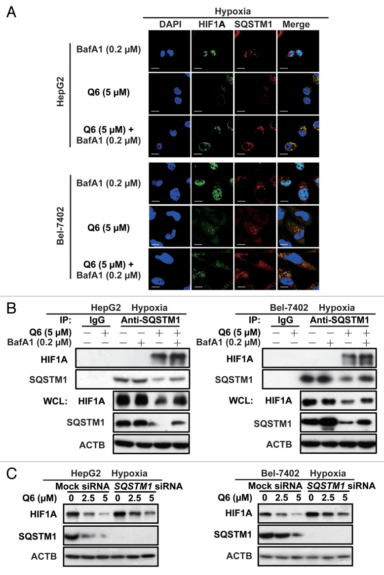
Tumor hypoxia underlies treatment failure and yields more aggressive and metastatic cancer phenotypes. Although therapeutically targeting these hypoxic environments has been proposed for many years, to date no approaches have shown the therapeutic value to gain regulatory approval. Here, we demonstrated that a novel hypoxia-activated prodrug, Q6, exhibits potent antiproliferative efficacy under hypoxic conditions and induces caspase-dependent apoptosis in 2 hepatocellular carcinoma (HCC) cell lines, with no obvious toxicity being detected in 2 normal liver cell lines. Treatment with Q6 markedly downregulated HIF1A [hypoxia inducible factor 1, α subunit (basic helix-loop-helix transcription factor)] expression and transcription of the downstream target gene, VEGFA (vascular endothelial growth factor A). This dual hypoxia-targeted modulation mechanism leads to high potency in suppressing tumor growth and vascularization in 2 in vivo models. Intriguingly, it is the autophagy-dependent degradation pathway that plays a crucial role in Q6-induced attenuation of HIF1A expression, rather than the proteasome-dependent pathway, which is normally regarded as the predominant mechanism underlying posttranslational regulation of HIF1A. Inhibition of autophagy, either by short interfering RNA (siRNA) or by chemical inhibitors, blocked Q6-induced HIF1A degradation. Autophagic degradation of HIF1A was further confirmed by the observation that HIF1A coimmunoprecipitated with the ubiquitin-binding adaptor protein, SQSTM1, which is degraded through autophagy. Additionally, silencing of SQSTM1 inhibited Q6-induced HIF1A degradation. These findings suggest that the novel hypoxia-targeted agent, Q6, has potential clinical value in the therapy of HCC. Furthermore, the identification of autophagy as a crucial regulator of HIF1A provides new insights into hypoxia-related treatments.
肿瘤缺氧是治疗失败的原因,并产生更具侵袭性和转移性的癌症表型。尽管多年来一直有人提出针对这些缺氧环境进行治疗,但迄今为止,尚无方法显示出具有获得监管批准的治疗价值。在此,我们证明了一种新型的缺氧激活前药Q6在缺氧条件下具有强大的抗增殖功效,并在2种肝癌(HCC)细胞系中诱导了半胱天冬酶依赖性凋亡,而在2种正常肝细胞系中未检测到明显毒性。用Q6处理可显著下调HIF1A[缺氧诱导因子1,α亚基(碱性螺旋-环-螺旋转录因子)]的表达以及下游靶基因VEGFA(血管内皮生长因子A)的转录。这种双重缺氧靶向调节机制在2种体内模型中具有高效抑制肿瘤生长和血管生成的作用。有趣的是,在Q6诱导的HIF1A表达减弱中起关键作用的是自噬依赖性降解途径,而非蛋白酶体依赖性途径,蛋白酶体依赖性途径通常被视为HIF1A翻译后调控的主要机制。通过短发夹RNA(shRNA)或化学抑制剂抑制自噬,均可阻断Q6诱导的HIF1A降解。HIF1A与泛素结合衔接蛋白SQSTM1共免疫沉淀,而SQSTM1可通过自噬降解,这一观察结果进一步证实了HIF1A的自噬性降解。此外,沉默SQSTM1可抑制Q6诱导的HIF1A降解。这些发现表明,新型缺氧靶向药物Q6在HCC治疗中具有潜在的临床价值。此外,将自噬鉴定为HIF1A的关键调节因子,为缺氧相关治疗提供了新的见解。