Hagenlocher Cathrin, Walentek Peter, M Ller Christina, Thumberger Thomas, Feistel Kerstin
Institute of Zoology, University of Hohenheim, Garbenstr, 30, Stuttgart 70593, Germany.
Cilia. 2013 Sep 24;2(1):12. doi: 10.1186/2046-2530-2-12.
Circulation of cerebrospinal fluid (CSF) through the ventricular system is driven by motile cilia on ependymal cells of the brain. Disturbed ciliary motility induces the formation of hydrocephalus, a pathological accumulation of CSF resulting in ventricle dilatation and increased intracranial pressure. The mechanism by which loss of motile cilia causes hydrocephalus has not been elucidated. The aim of this study was: (1) to provide a detailed account of the development of ciliation in the brain of the African clawed frog Xenopus laevis; and (2) to analyze the relevance of ependymal cilia motility for CSF circulation and brain ventricle morphogenesis in Xenopus.
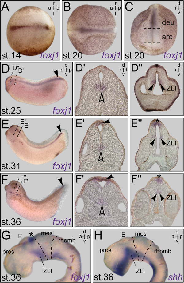
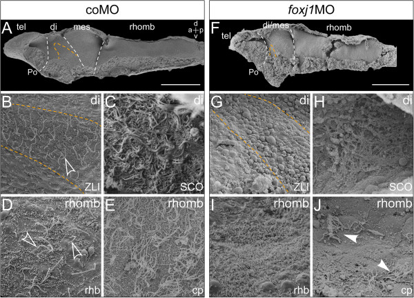
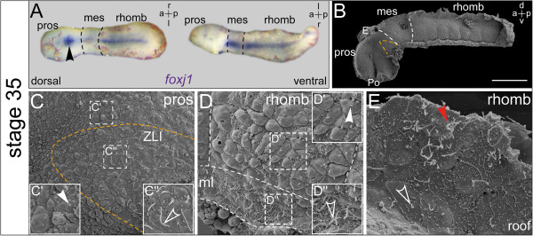
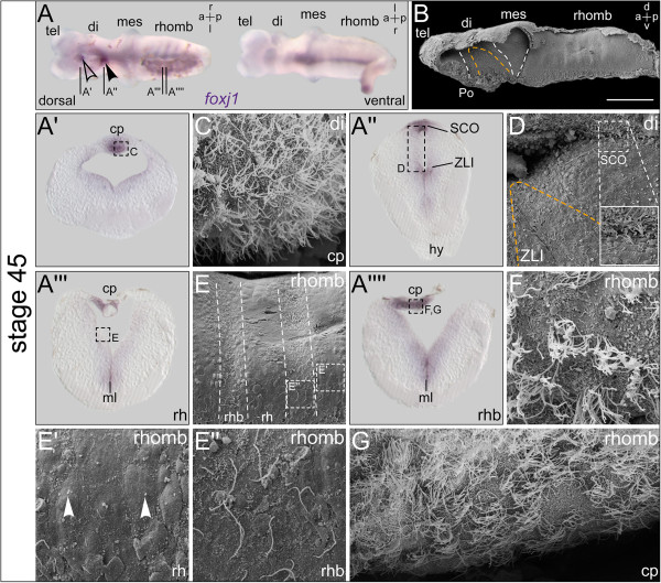
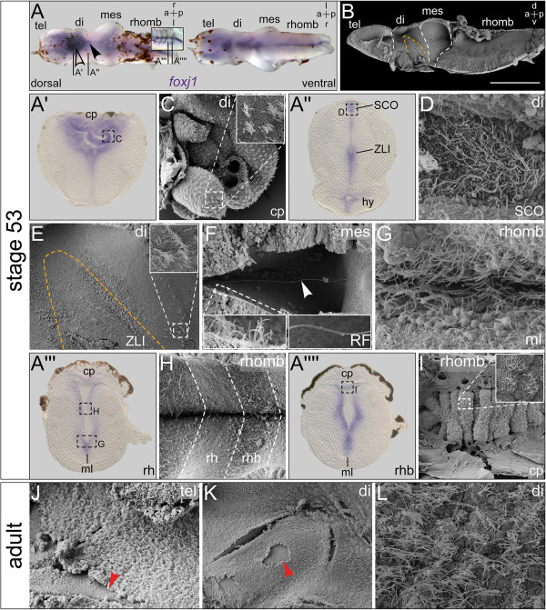
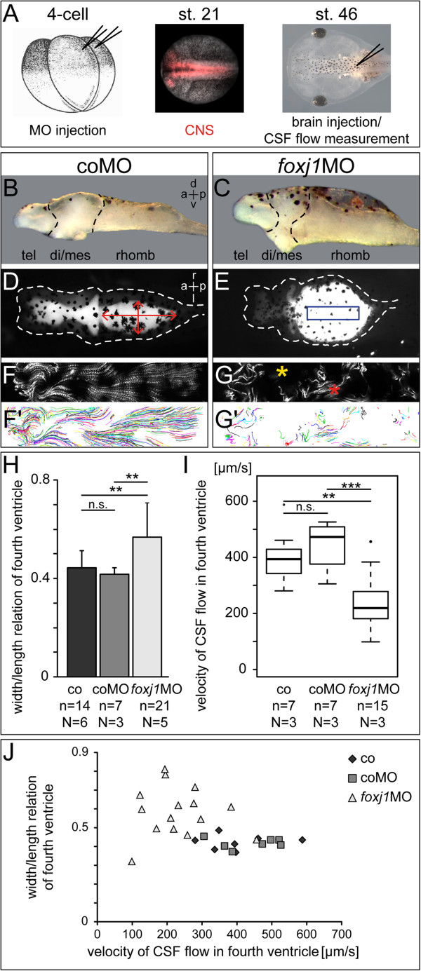
Gene expression analysis of foxj1, the bona fide marker for motile cilia, was used to identify potentially ciliated regions in the developing central nervous system (CNS) of the tadpole. Scanning electron microscopy (SEM) was used to reveal the distribution of mono- and multiciliated cells during successive stages of brain morphogenesis, which was functionally assessed by bead injection and video microscopy of ventricular CSF flow. An antisense morpholino oligonucleotide (MO)-mediated gene knock-down that targeted foxj1 in the CNS was applied to assess the role of motile cilia in the ventricles.
RNA transcripts of foxj1 in the CNS were found from neurula stages onwards. Following neural tube closure, foxj1 expression was seen in distinct ventricular regions such as the zona limitans intrathalamica (ZLI), subcommissural organ (SCO), floor plate, choroid plexus (CP), and rhombomere boundaries. In all areas, expression of foxj1 preceded the outgrowth of monocilia and the subsequent switch to multiciliated ependymal cells. Cilia were absent in foxj1 morphants, causing impaired CSF flow and fourth ventricle hydrocephalus in tadpole-stage embryos.
Motile ependymal cilia are important organelles in the Xenopus CNS, as they are essential for the circulation of CSF and maintenance of homeostatic fluid pressure. The Xenopus CNS ventricles might serve as a novel model system for the analysis of human ciliary genes whose deficiency cause hydrocephalus.
脑脊液(CSF)在脑室系统中的循环由脑室内室管膜细胞上的运动性纤毛驱动。纤毛运动障碍会导致脑积水的形成,脑积水是脑脊液的病理性积聚,导致脑室扩张和颅内压升高。运动性纤毛丧失导致脑积水的机制尚未阐明。本研究的目的是:(1)详细描述非洲爪蟾(Xenopus laevis)脑中纤毛形成的发育过程;(2)分析室管膜纤毛运动对非洲爪蟾脑脊液循环和脑室形态发生的相关性。
使用运动性纤毛的真正标志物foxj1的基因表达分析来鉴定蝌蚪发育中的中枢神经系统(CNS)中潜在的纤毛区域。扫描电子显微镜(SEM)用于揭示脑形态发生连续阶段中单纤毛和多纤毛细胞的分布,并通过珠子注射和脑室脑脊液流动的视频显微镜对其功能进行评估。应用针对中枢神经系统中foxj1的反义吗啉代寡核苷酸(MO)介导的基因敲低来评估运动性纤毛在脑室中的作用。
从神经胚阶段开始就在中枢神经系统中发现了foxj1的RNA转录本。神经管闭合后,在不同的脑室区域观察到foxj1表达,如丘脑间界限带(ZLI)、联合下器官(SCO)、底板、脉络丛(CP)和菱脑节边界。在所有区域,foxj1的表达先于单纤毛的长出以及随后向多纤毛室管膜细胞的转变。foxj1基因敲降的胚胎中没有纤毛,导致蝌蚪期胚胎的脑脊液流动受损和第四脑室脑积水。
运动性室管膜纤毛是非洲爪蟾中枢神经系统中的重要细胞器,因为它们对于脑脊液循环和维持稳态流体压力至关重要。非洲爪蟾中枢神经系统脑室可能作为一种新型模型系统,用于分析其缺陷导致脑积水的人类纤毛基因。