• 文献检索
  • 文档翻译
  • 深度研究
  • 学术资讯
  • Suppr Zotero 插件Zotero 插件
  • 邀请有礼
  • 套餐&价格
  • 历史记录
应用&插件
Suppr Zotero 插件Zotero 插件浏览器插件Mac 客户端Windows 客户端微信小程序
定价
高级版会员购买积分包购买API积分包
服务
文献检索文档翻译深度研究API 文档MCP 服务
关于我们
关于 Suppr公司介绍联系我们用户协议隐私条款
关注我们

Suppr 超能文献

核心技术专利:CN118964589B侵权必究
粤ICP备2023148730 号-1Suppr @ 2026

文献检索

告别复杂PubMed语法,用中文像聊天一样搜索,搜遍4000万医学文献。AI智能推荐,让科研检索更轻松。

立即免费搜索

文件翻译

保留排版,准确专业,支持PDF/Word/PPT等文件格式,支持 12+语言互译。

免费翻译文档

深度研究

AI帮你快速写综述,25分钟生成高质量综述,智能提取关键信息,辅助科研写作。

立即免费体验

一种用于评估青光眼患者神经视网膜边缘组织量的方法:与目前用于测量边缘面积的方法比较。

A method to estimate the amount of neuroretinal rim tissue in glaucoma: comparison with current methods for measuring rim area.

机构信息

Devers Eye Institute, Legacy Research Institute, Portland, Oregon.

Devers Eye Institute, Legacy Research Institute, Portland, Oregon; New York Eye and Ear Infirmary, New York, New York.

出版信息

Am J Ophthalmol. 2014 Mar;157(3):540-9.e1-2. doi: 10.1016/j.ajo.2013.11.007. Epub 2013 Nov 13.

DOI:10.1016/j.ajo.2013.11.007
PMID:24239775
原文链接:https://pmc.ncbi.nlm.nih.gov/articles/PMC3944716/
Abstract

PURPOSE

To test whether the minimum rim area assessed by spectral domain optical coherence tomography (SD-OCT), based on the shortest distance from the Bruch membrane opening (BMO) to the inner limiting membrane, corresponds more closely to retinal nerve fiber layer (RNFL) thickness and visual field mean deviation (MD) than current rim measures in early glaucoma.

DESIGN

Prospective cross-sectional study.

METHODS

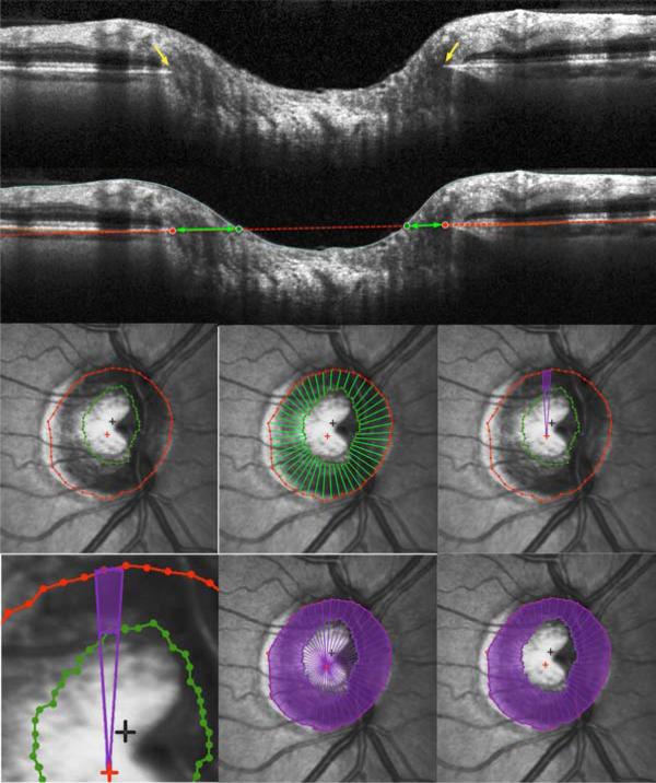
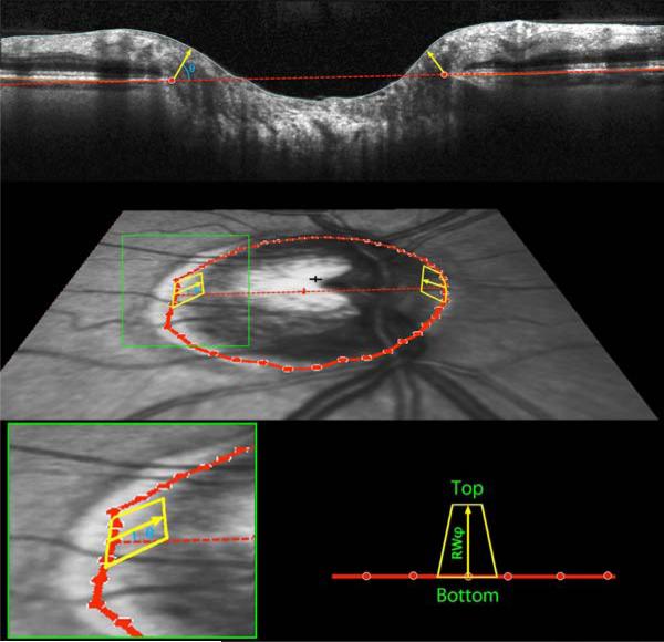
We studied 221 participants with non-endstage glaucoma or high-risk ocular hypertension and performed standard automated perimetry. We received SD-OCT and confocal scanning laser ophthalmoscopy (CSLO) scans on the same day. Rim area measured by CSLO was compared with 3 SD-OCT rim measures from radial B-scans: horizontal rim area between BMO and inner limiting membrane within the BMO plane; mean minimum rim width (BMO-MRW); and minimum rim area (BMO-MRA) optimized within sectors and then summed. Correlations between these measures and either MD from perimetry or RNFL thickness from SD-OCT were compared using the Steiger test.

RESULTS

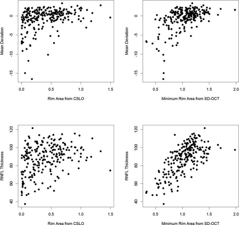
RNFL thickness was better correlated with BMO-MRA (r = 0.676) or BMO-MRW (r = 0.680) than with either CSLO rim area (r = 0.330, P < 0.001) or horizontal rim area (r = 0.482, P < 0.001). MD was better correlated with BMO-MRA (r = 0.534) or BMO-MRW (r = 0.546) than with either CSLO rim area (r = 0.321, P < 0.001) or horizontal rim area (0.403, P < 0.001). The correlation between MD and RNFL thickness was r = 0.646.

CONCLUSIONS

Minimum rim measurements from SD-OCT are significantly better correlated to both RNFL thickness and MD than rim measurements within the BMO plane or based on the clinical disc margin. They provide new structural parameters for both diagnostic and research purposes in glaucoma.

摘要

目的

通过评估基于从 Bruch 膜开口(BMO)到内界膜最短距离的光谱域光相干断层扫描(SD-OCT)最小边缘区域,来检测其是否比当前早期青光眼边缘测量值更能准确反映视网膜神经纤维层(RNFL)厚度和视野平均偏差(MD)。

设计

前瞻性横断面研究。

方法

我们研究了 221 名非晚期青光眼或高危眼高压患者,并进行了标准自动视野检查。同一天我们进行了 SD-OCT 和共焦扫描激光检眼镜(CSLO)扫描。CSLO 测量的边缘区域与来自径向 B 扫描的 3 个 SD-OCT 边缘区域进行了比较:BMO 平面内 BMO 和内界膜之间的水平边缘区域;平均最小边缘宽度(BMO-MRW);以及在扇区中进行优化后的最小边缘区域(BMO-MRA),然后对其进行求和。使用 Steiger 检验比较这些测量值与视野 MD 或 SD-OCT 中 RNFL 厚度之间的相关性。

结果

与 CSLO 边缘区域(r = 0.330,P < 0.001)或水平边缘区域(r = 0.482,P < 0.001)相比,RNFL 厚度与 BMO-MRA(r = 0.676)或 BMO-MRW(r = 0.680)相关性更好。MD 与 BMO-MRA(r = 0.534)或 BMO-MRW(r = 0.546)相关性更好,与 CSLO 边缘区域(r = 0.321,P < 0.001)或水平边缘区域(0.403,P < 0.001)相关性较差。MD 和 RNFL 厚度之间的相关性为 r = 0.646。

结论

与 BMO 平面内或基于临床视盘边缘的边缘测量值相比,SD-OCT 的最小边缘测量值与 RNFL 厚度和 MD 的相关性显著更高。它们为青光眼的诊断和研究目的提供了新的结构参数。

https://cdn.ncbi.nlm.nih.gov/pmc/blobs/15b7/3944716/ecce2f0c50fb/nihms-540854-f0004.jpg
https://cdn.ncbi.nlm.nih.gov/pmc/blobs/15b7/3944716/ecc856f34207/nihms-540854-f0002.jpg
https://cdn.ncbi.nlm.nih.gov/pmc/blobs/15b7/3944716/07f62beb0a7f/nihms-540854-f0003.jpg
https://cdn.ncbi.nlm.nih.gov/pmc/blobs/15b7/3944716/ecce2f0c50fb/nihms-540854-f0004.jpg
https://cdn.ncbi.nlm.nih.gov/pmc/blobs/15b7/3944716/ecc856f34207/nihms-540854-f0002.jpg
https://cdn.ncbi.nlm.nih.gov/pmc/blobs/15b7/3944716/07f62beb0a7f/nihms-540854-f0003.jpg
https://cdn.ncbi.nlm.nih.gov/pmc/blobs/15b7/3944716/ecce2f0c50fb/nihms-540854-f0004.jpg

相似文献

1
A method to estimate the amount of neuroretinal rim tissue in glaucoma: comparison with current methods for measuring rim area.一种用于评估青光眼患者神经视网膜边缘组织量的方法:与目前用于测量边缘面积的方法比较。
Am J Ophthalmol. 2014 Mar;157(3):540-9.e1-2. doi: 10.1016/j.ajo.2013.11.007. Epub 2013 Nov 13.
2
Novel Bruch's Membrane Opening Minimum Rim Area Equalizes Disc Size Dependency and Offers High Diagnostic Power for Glaucoma.新型布鲁赫膜开口最小边缘面积可平衡视盘大小依赖性并为青光眼提供高诊断效能。
Invest Ophthalmol Vis Sci. 2016 Dec 1;57(15):6596-6603. doi: 10.1167/iovs.16-20561.
3
Evaluation of two-dimensional Bruch's membrane opening minimum rim area for glaucoma diagnostics in a large patient cohort.大样本患者群体中二维 Bruch 膜开口最小边缘区域在青光眼诊断中的评估。
Acta Ophthalmol. 2019 Feb;97(1):60-67. doi: 10.1111/aos.13698. Epub 2018 Mar 24.
4
Diagnostic Accuracy of Optical Coherence Tomography and Scanning Laser Tomography for Identifying Glaucoma in Myopic Eyes.光学相干断层扫描和扫描激光断层扫描在识别近视眼中青光眼的诊断准确性。
Ophthalmology. 2016 Jun;123(6):1181-9. doi: 10.1016/j.ophtha.2016.01.052. Epub 2016 Mar 15.
5
Structure-function relationships with spectral-domain optical coherence tomography retinal nerve fiber layer and optic nerve head measurements.光谱域光学相干断层扫描视网膜神经纤维层和视神经乳头测量的结构-功能关系
Invest Ophthalmol Vis Sci. 2014 May 2;55(5):2953-62. doi: 10.1167/iovs.13-13482.
6
Enhanced detection of open-angle glaucoma with an anatomically accurate optical coherence tomography-derived neuroretinal rim parameter.利用解剖精确的光学相干断层扫描衍生的神经视网膜边缘参数增强开角型青光眼的检测。
Ophthalmology. 2013 Mar;120(3):535-543. doi: 10.1016/j.ophtha.2012.09.055. Epub 2012 Dec 23.
7
Enhanced structure-function relationship in glaucoma with an anatomically and geometrically accurate neuroretinal rim measurement.通过解剖学和几何学精确的神经视网膜边缘测量,青光眼患者的结构-功能关系得到增强。
Invest Ophthalmol Vis Sci. 2014 Dec 11;56(1):98-105. doi: 10.1167/iovs.14-15375.
8
The use of Bruch's membrane opening-based optical coherence tomography of the optic nerve head for glaucoma detection in microdiscs.基于布鲁赫膜开口的视神经乳头光学相干断层扫描技术在微小视盘中检测青光眼的应用。
Br J Ophthalmol. 2017 Apr;101(4):530-535. doi: 10.1136/bjophthalmol-2016-308957. Epub 2016 Jul 19.
9
Effect of Trabeculectomy on OCT Measurements of the Optic Nerve Head Neuroretinal Rim Tissue.小梁切除术对 OCT 测量视神经头神经视网膜边缘组织的影响。
Ophthalmol Glaucoma. 2020 Jan-Feb;3(1):32-39. doi: 10.1016/j.ogla.2019.09.003. Epub 2019 Oct 4.
10
Neuroretinal rim in non-glaucomatous large optic nerve heads: a comparison of confocal scanning laser tomography and spectral domain optical coherence tomography.非青光眼性大视乳头的神经视网膜边缘:共焦扫描激光断层扫描与光谱域光学相干断层扫描的比较
Br J Ophthalmol. 2017 Feb;101(2):138-142. doi: 10.1136/bjophthalmol-2015-307730. Epub 2016 Apr 26.

引用本文的文献

1
[Progression assessment of glaucoma].[青光眼的病情进展评估]
Ophthalmologie. 2025 Sep 4. doi: 10.1007/s00347-025-02302-x.
2
Spectralis Optical Coherence Tomography for Evaluating Ocular Hypertensive and Glaucoma Suspect Eyes: Real-World Data from Taiwan.使用Spectralis光学相干断层扫描技术评估高眼压症和疑似青光眼患者的眼部:来自台湾的真实世界数据
Diagnostics (Basel). 2025 May 15;15(10):1256. doi: 10.3390/diagnostics15101256.
3
Interpretable Machine Learning Predictions of Bruch's Membrane Opening-Minimum Rim Width Using Retinal Nerve Fiber Layer Values and Visual Field Global Indexes.

本文引用的文献

1
From clinical examination of the optic disc to clinical assessment of the optic nerve head: a paradigm change.从视盘临床检查到视乳头临床评估:范式转变。
Am J Ophthalmol. 2013 Aug;156(2):218-227.e2. doi: 10.1016/j.ajo.2013.04.016. Epub 2013 Jun 12.
2
Enhanced detection of open-angle glaucoma with an anatomically accurate optical coherence tomography-derived neuroretinal rim parameter.利用解剖精确的光学相干断层扫描衍生的神经视网膜边缘参数增强开角型青光眼的检测。
Ophthalmology. 2013 Mar;120(3):535-543. doi: 10.1016/j.ophtha.2012.09.055. Epub 2012 Dec 23.
3
Relationship between orbital optic nerve axon counts and retinal nerve fiber layer thickness measured by spectral domain optical coherence tomography.
使用视网膜神经纤维层值和视野全局指标对布鲁赫膜开口-最小边缘宽度进行可解释的机器学习预测。
Bioengineering (Basel). 2025 Mar 20;12(3):321. doi: 10.3390/bioengineering12030321.
4
Reversal of cup-to-disc ratio following trabeculectomy using optical coherence tomography imaging: A case series.使用光学相干断层扫描成像观察小梁切除术后杯盘比的逆转:病例系列
Saudi J Ophthalmol. 2024 Dec 2;38(4):391-395. doi: 10.4103/sjopt.sjopt_165_24. eCollection 2024 Oct-Dec.
5
[Visual field prediction based on temporal-spatial feature learning].基于时空特征学习的视野预测
Sheng Wu Yi Xue Gong Cheng Xue Za Zhi. 2024 Oct 25;41(5):1003-1011. doi: 10.7507/1001-5515.202310072.
6
Acute effects of swimming goggle wearing on intraocular pressure, anterior chamber biometrics, and optic nerve head morphology.游泳镜佩戴对眼压、前房生物测量和视乳头形态的急性影响。
Sci Rep. 2024 Oct 7;14(1):23339. doi: 10.1038/s41598-024-73990-9.
7
Unveiling Novel Structural Biomarkers for the Diagnosis of Glaucoma.揭示用于青光眼诊断的新型结构生物标志物。
Biomedicines. 2024 May 29;12(6):1211. doi: 10.3390/biomedicines12061211.
8
Intraday repeatability of macular layers measurements in glaucomatous and non-glaucomatous patients using spectral-domain optical coherence tomography.利用频域光学相干断层扫描测量青光眼和非青光眼患者的黄斑层重复性。
Graefes Arch Clin Exp Ophthalmol. 2024 Oct;262(10):3287-3294. doi: 10.1007/s00417-024-06498-7. Epub 2024 May 6.
9
The Temporal Relation Between Rates of Retinal Nerve Fiber Layer and Minimum Rim Width Changes in Glaucoma.青光眼的视网膜神经纤维层和最小视盘边缘宽度变化率之间的时间关系。
Transl Vis Sci Technol. 2024 Apr 2;13(4):10. doi: 10.1167/tvst.13.4.10.
10
Morphological differences of the neuroretinal rim between temporally tilted and non-tilted optic discs in healthy eyes.健康眼中视盘倾斜与非倾斜时神经视网膜边缘的形态差异。
Sci Rep. 2024 Mar 13;14(1):6070. doi: 10.1038/s41598-024-54116-7.
通过谱域光学相干断层扫描测量的眶内视神经轴突计数与视网膜神经纤维层厚度的关系。
Invest Ophthalmol Vis Sci. 2012 Nov 21;53(12):7766-73. doi: 10.1167/iovs.12-10752.
4
Measurement of optic disc size and rim area with spectral-domain OCT and scanning laser ophthalmoscopy.使用频域光学相干断层扫描和扫描激光检眼镜测量视盘大小和边缘面积。
Invest Ophthalmol Vis Sci. 2012 Jul 9;53(8):4519-30. doi: 10.1167/iovs.11-8362.
5
Factors predicting the rate of functional progression in early and suspected glaucoma.预测早期和疑似青光眼功能性进展速度的因素。
Invest Ophthalmol Vis Sci. 2012 Jun 14;53(7):3598-604. doi: 10.1167/iovs.11-9065.
6
The effect of test variability on the structure-function relationship in early glaucoma.测试变异性对早期青光眼结构-功能关系的影响。
Graefes Arch Clin Exp Ophthalmol. 2012 Dec;250(12):1851-61. doi: 10.1007/s00417-012-2005-9. Epub 2012 Apr 25.
7
Structure-function relationships between spectral-domain OCT and standard achromatic perimetry.频域光学相干断层扫描与标准消色差视野检查的结构-功能关系。
Invest Ophthalmol Vis Sci. 2012 May 9;53(6):2740-8. doi: 10.1167/iovs.11-8320.
8
Influence of clinically invisible, but optical coherence tomography detected, optic disc margin anatomy on neuroretinal rim evaluation.临床不可见但光相干断层扫描可检测到的视盘边缘解剖结构对神经视网膜边缘评估的影响。
Invest Ophthalmol Vis Sci. 2012 Apr 18;53(4):1852-60. doi: 10.1167/iovs.11-9309.
9
Combining structural and functional measurements to improve estimates of rates of glaucomatous progression.结合结构和功能测量来提高青光眼进展速度的估计值。
Am J Ophthalmol. 2012 Jun;153(6):1197-205.e1. doi: 10.1016/j.ajo.2011.11.015. Epub 2012 Feb 7.
10
Optic disc margin anatomy in patients with glaucoma and normal controls with spectral domain optical coherence tomography.利用频域光相干断层扫描技术观察青光眼患者和正常对照者视盘边缘解剖结构。
Ophthalmology. 2012 Apr;119(4):738-47. doi: 10.1016/j.ophtha.2011.09.054. Epub 2012 Jan 4.