Departments of Psychiatry, Washington University School of Medicine, St, Louis, MO 63110, USA.
Acta Neuropathol Commun. 2013 Jun 12;1:23. doi: 10.1186/2051-5960-1-23.
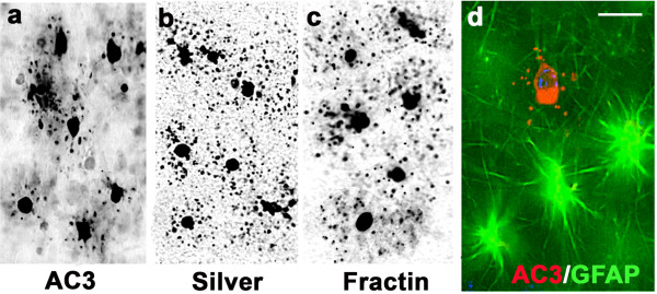
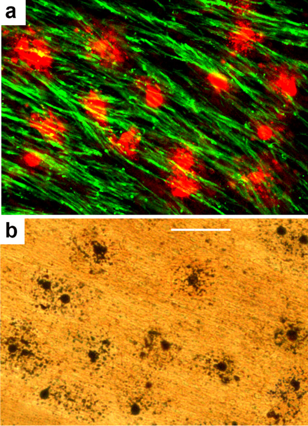
In utero exposure of the fetal non-human primate (NHP) brain to alcohol on a single occasion during early or late third-trimester gestation triggers widespread acute apoptotic death of cells in both gray and white matter (WM) regions of the fetal brain. In a prior publication, we documented that the dying gray matter cells are neurons, and described the regional distribution and magnitude of this cell death response. Here, we present new findings regarding the magnitude, identity and maturational status of the dying WM cells in these alcohol-exposed fetal NHP brains.
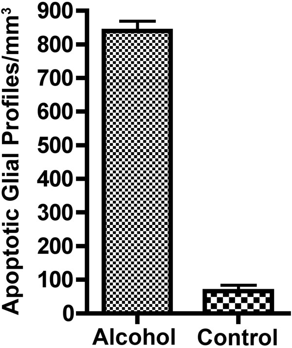
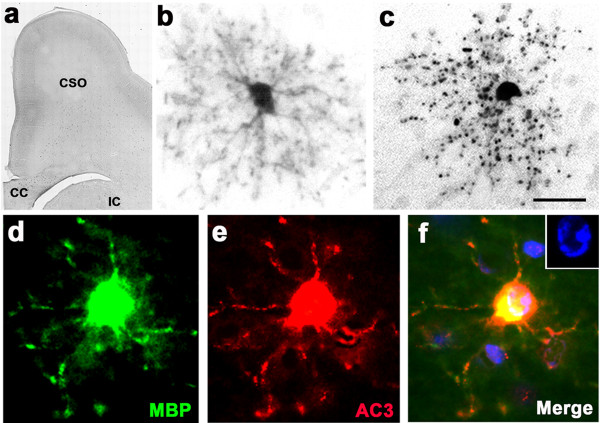
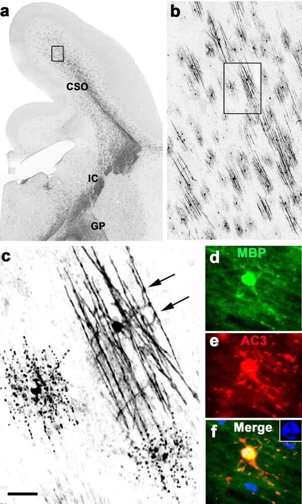
Our findings document that the dying WM cells belong to the oligodendrocyte (OL) lineage. OLs become vulnerable when they are just beginning to generate myelin basic protein in preparation for myelinating axons, and they remain vulnerable throughout later stages of myelination. We found no evidence linking astrocytes, microglia or OL progenitors to this WM cell death response. The mean density (profiles per mm3) of dying WM cells in alcohol-exposed brains was 12.7 times higher than the mean density of WM cells dying by natural apoptosis in drug-naive control brains.
In utero exposure of the fetal NHP brain to alcohol on a single occasion triggers widespread acute apoptotic death of neurons (previous study) and of OLs (present study) throughout WM regions of the developing brain. The rate of OL apoptosis in alcohol-exposed brains was 12.7 times higher than the natural OL apoptosis rate. OLs become sensitive to the apoptogenic action of alcohol when they are just beginning to generate constituents of myelin in their cytoplasm, and they remain vulnerable throughout later stages of myelination. There is growing evidence for a similar apoptotic response of both neurons and OLs following exposure of the developing brain to anesthetic and anticonvulsant drugs. Collectively, this body of evidence raises important questions regarding the role that neuro and oligo apoptosis may play in the human condition known as fetal alcohol spectrum disorder (FASD), and also poses a question whether other apoptogenic drugs, although long considered safe for pediatric/obstetric use, may have the potential to cause iatrogenic FASD-like developmental disability syndromes.
在妊娠晚期的单个第三孕期,胎儿灵长类动物(NHP)的大脑单次暴露于酒精会引发胎儿大脑的灰质和白质(WM)区域中广泛的急性细胞凋亡死亡。在之前的一篇出版物中,我们记录了死亡的灰质细胞是神经元,并描述了这种细胞死亡反应的区域分布和程度。在这里,我们提出了关于这些暴露于酒精的胎儿 NHP 大脑中死亡 WM 细胞的数量、身份和成熟状态的新发现。
我们的研究结果表明,死亡的 WM 细胞属于少突胶质细胞(OL)谱系。OL 变得脆弱,当它们刚刚开始产生髓鞘碱性蛋白,为髓鞘形成轴突做准备时,并且在髓鞘形成的后期阶段仍然脆弱。我们没有发现将星形胶质细胞、小胶质细胞或 OL 祖细胞与这种 WM 细胞死亡反应联系起来的证据。暴露于酒精的大脑中死亡 WM 细胞的平均密度(每立方毫米的剖面)比药物-naive 对照大脑中自然凋亡的 WM 细胞死亡的平均密度高 12.7 倍。
在单个第三孕期,胎儿 NHP 的大脑单次暴露于酒精会引发整个 WM 区域的神经元(之前的研究)和 OL(目前的研究)的广泛急性凋亡死亡。暴露于酒精的大脑中 OL 的凋亡率比自然 OL 凋亡率高 12.7 倍。当 OL 开始在细胞质中产生髓鞘成分时,它们对酒精的促凋亡作用变得敏感,并且在髓鞘形成的后期阶段仍然脆弱。越来越多的证据表明,麻醉和抗惊厥药物暴露于发育中的大脑后,神经元和 OL 都有类似的凋亡反应。总的来说,这一系列证据提出了一个重要的问题,即神经和 OL 凋亡在被称为胎儿酒精谱系障碍(FASD)的人类疾病中可能起什么作用,并提出了一个问题,即尽管长期以来被认为对儿科/产科使用安全的其他促凋亡药物是否有可能导致医源性 FASD 样发育障碍综合征。