Department of Pediatrics, Carver College of Medicine, University of Iowa, Iowa City, IA 2524 JCP, USA.
Acta Neuropathol Commun. 2013 Oct 10;1:66. doi: 10.1186/2051-5960-1-66.
Medulloblastoma is the most common malignant brain tumor in children. Genetic profiling has identified four principle tumor subgroups; each subgroup is characterized by different initiating mutations, genetic and clinical profiles, and prognoses. The two most well-defined subgroups are caused by overactive signaling in the WNT and SHH mitogenic pathways; less is understood about Groups 3 and 4 medulloblastoma. Identification of tumor subgroup using molecular classification is set to become an important component of medulloblastoma diagnosis and staging, and will likely guide therapeutic options. However, thus far, few druggable targets have emerged. G-protein coupled receptors (GPCRs) possess characteristics that make them ideal targets for molecular imaging and therapeutics; drugs targeting GPCRs account for 30-40% of all current pharmaceuticals. While expression patterns of many proteins in human medulloblastoma subgroups have been discerned, the expression pattern of GPCRs in medulloblastoma has not been investigated. We hypothesized that analysis of GPCR expression would identify clear subsets of medulloblastoma and suggest distinct GPCRs that might serve as molecular targets for both imaging and therapy.
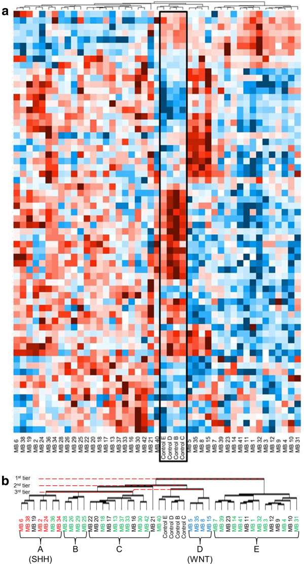
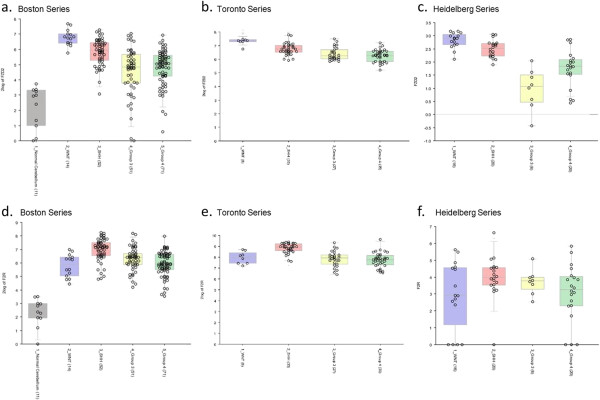
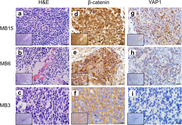
Our study found that medulloblastoma tumors fall into distinct clusters based solely on GPCR expression patterns. Normal cerebellum clustered separately from the tumor samples. Further, two of the tumor clusters correspond with high fidelity to the WNT and SHH subgroups of medulloblastoma. Distinct over-expressed GPCRs emerge; for example, LGR5 and GPR64 are significantly and uniquely over-expressed in the WNT subgroup of tumors, while PTGER4 is over-expressed in the SHH subgroup. Uniquely under-expressed GPCRs were also observed. Our key findings were independently validated using a large international dataset.
Our results identify GPCRs with potential to act as imaging and therapeutic targets. Elucidating tumorigenic pathways is a secondary benefit to identifying differential GPCR expression patterns in medulloblastoma tumors.
成神经管细胞瘤是儿童中最常见的恶性脑肿瘤。基因谱分析确定了四个主要的肿瘤亚组;每个亚组的特征是不同的起始突变、遗传和临床特征以及预后。两个最明确的亚组是由 WNT 和 SHH 有丝分裂信号通路的过度激活引起的;对第 3 组和第 4 组成神经管细胞瘤的了解较少。使用分子分类鉴定肿瘤亚组将成为成神经管细胞瘤诊断和分期的重要组成部分,并且可能指导治疗选择。然而,迄今为止,很少有可靶向的靶点出现。G 蛋白偶联受体 (GPCR) 具有使其成为分子成像和治疗的理想靶点的特征;靶向 GPCR 的药物占所有现有药物的 30-40%。虽然已经发现了人类成神经管细胞瘤亚组中许多蛋白质的表达模式,但成神经管细胞瘤中 GPCR 的表达模式尚未被研究。我们假设 GPCR 表达的分析将确定成神经管细胞瘤的明确亚组,并提出可能作为成像和治疗的分子靶标的不同 GPCR。
我们的研究发现,成神经管细胞瘤肿瘤仅根据 GPCR 表达模式分为不同的簇。正常小脑与肿瘤样本分开聚集。此外,两个肿瘤簇与成神经管细胞瘤的 WNT 和 SHH 亚组高度一致。出现明显过表达的 GPCR;例如,LGR5 和 GPR64 在 WNT 亚组的肿瘤中显著且独特地过表达,而 PTGER4 在 SHH 亚组中过表达。还观察到独特的低表达 GPCR。我们的主要发现使用大型国际数据集进行了独立验证。
我们的结果确定了具有作为成像和治疗靶标的潜力的 GPCR。阐明肿瘤发生途径是识别成神经管细胞瘤肿瘤中差异 GPCR 表达模式的次要益处。