Chu Hyun Hee, Hwang Pyoung Han, Jeong Yeon Jun, Chung Myoung Ja
Department of Pathology, Chonbuk National University Medical School, Jeonju, Korea.
Korean J Pathol. 2013 Oct;47(5):472-6. doi: 10.4132/KoreanJPathol.2013.47.5.472. Epub 2013 Oct 25.
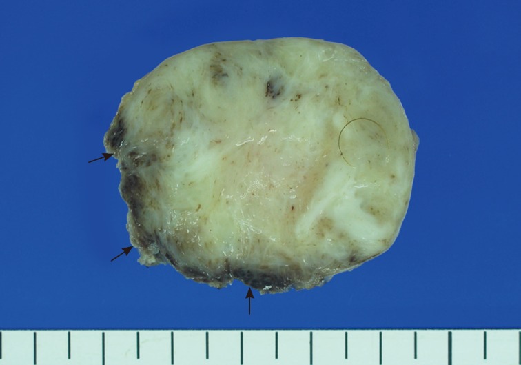
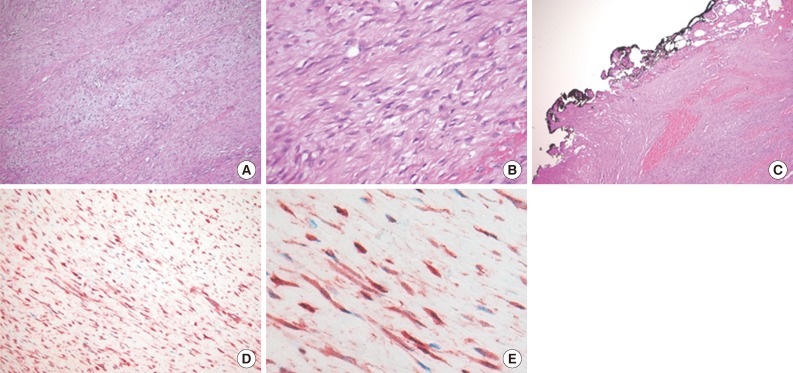
Fibromatoses comprise many different entities of well-differentiated fibroblastic proliferation with variable collagen production and form a firm nodular mass. Abdominal fibromatosis is distinguishable from other forms of fibromatosis because of its location and its tendency to occur in women of childbearing age during or following pregnancy. Abdominal fibromatosis in children is an extremely rare condition. A 15-month-old boy presented with an abdominal wall mass that had recently increased in size. Mass excision was perfomed. The tumor was 4.3×4.1 cm and partly circumscribed. Histologically, the tumor was composed of parallel long fascicles of spindle-cells with a uniform appearance. The edges of the resected mass were infiltrative, and the surgical margins were positive. Mitotic figures were <1/10 high power fields. No cellular atypia or necrosis was present. The tumor cells were positive for vimentin and nuclear β-catenin staining.
纤维瘤病由许多不同的、分化良好的成纤维细胞增殖实体组成,胶原产生情况各异,并形成坚实的结节状肿块。腹壁纤维瘤病因其位置以及在育龄期女性孕期或产后发病的倾向,有别于其他形式的纤维瘤病。儿童腹壁纤维瘤病极为罕见。一名15个月大的男童出现腹壁肿块,且肿块近期增大。进行了肿块切除术。肿瘤大小为4.3×4.1厘米,部分边界清晰。组织学上,肿瘤由外观一致的平行长梭形细胞束组成。切除肿块的边缘呈浸润性,手术切缘阳性。有丝分裂象<1/10高倍视野。未发现细胞异型性或坏死。肿瘤细胞波形蛋白和核β-连环蛋白染色呈阳性。