Deshwal Avinash, Walton Tessa, Varzgalis Manvydas, McGowan Katherine, O'Dowd Gerard
Department of Breast Surgery, Letterkenny University Hospital, County Donegal, Republic of Ireland.
Radiol Case Rep. 2023 Mar 17;18(5):1949-1953. doi: 10.1016/j.radcr.2023.02.019. eCollection 2023 May.
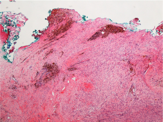
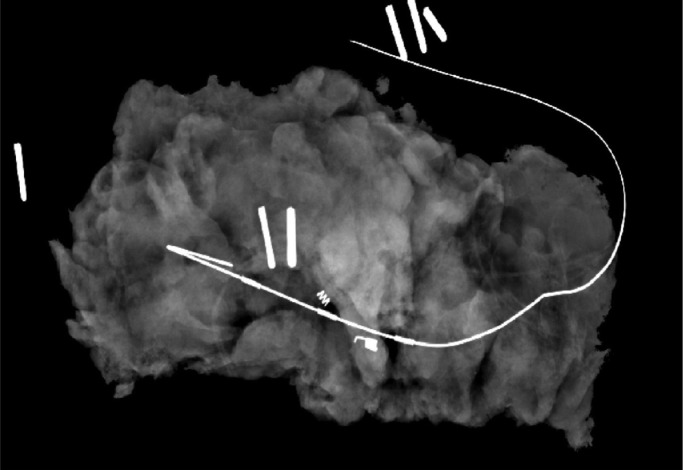
Mammary fibromatosis is a rare neoplastic proliferation of fibroblastic cells. Usually seen in abdominal and extra-abdominal sites, it is rarely seen in the breast. Patients with mammary fibromatosis usually present with a firm palpable mass with or without dimpling and skin retraction-often mimicking breast carcinoma. Here, we present a case of mammary fibromatosis in a 49-year-old woman who presented with a palpable lump in her right breast. Mammography tomosynthesis revealed architectural distortion which was seen on ultrasonography as a hypoechoic area. The patient underwent a wire-guided excision where the histology of this specimen showed irregular spindle cell proliferation with hemosiderin deposition, confirming mammary fibromatosis. Further re-excision of margins revealed no evidence of residual fibromatosis, and the patient underwent subsequent surveillance mammograms to ensure there was no recurrence.
乳腺纤维瘤病是一种罕见的成纤维细胞肿瘤性增生。通常见于腹部和腹外部位,很少见于乳腺。乳腺纤维瘤病患者通常表现为可触及的坚实肿块,伴有或不伴有酒窝征和皮肤凹陷,常类似乳腺癌。在此,我们报告一例49岁女性乳腺纤维瘤病病例,该患者右乳可触及肿块。乳腺断层合成摄影显示结构紊乱,超声检查显示为低回声区。患者接受了钢丝引导下切除,该标本的组织学检查显示不规则梭形细胞增生并伴有含铁血黄素沉积,确诊为乳腺纤维瘤病。进一步切除边缘组织未见残留纤维瘤病迹象,患者随后接受了乳腺钼靶复查以确保无复发。