1. Department of Biomedical Engineering, University of Michigan, Ann Arbor, MI, USA;
Theranostics. 2013 Oct 20;3(11):851-64. doi: 10.7150/thno.6717. eCollection 2013.
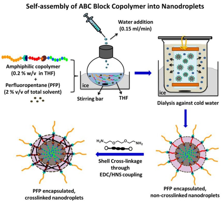
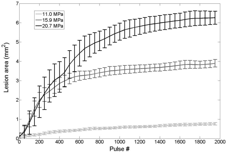
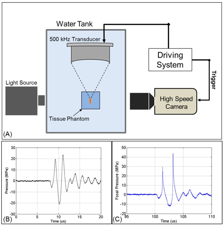
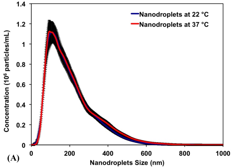
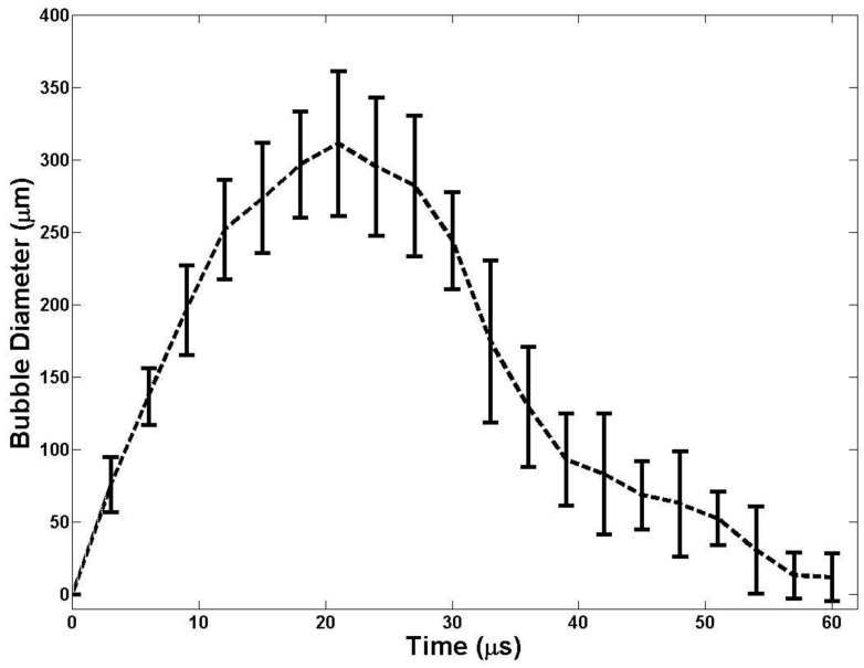
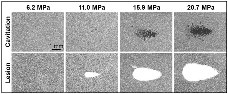
This paper is an initial work towards developing an image-guided, targeted ultrasound ablation technique by combining histotripsy with nanodroplets that can be selectively delivered to tumor cells. Using extremely short, high-pressure pulses, histotripsy generates a dense cloud of cavitating microbubbles that fractionates tissue. We hypothesize that synthetic nanodroplets that encapsulate a perfluoropentane (PFP) core will transition upon exposure to ultrasound pulses into gas microbubbles, which will rapidly expand and collapse resulting in disruption of cells similar to the histotripsy process but at a significantly lower acoustic pressure. The significantly reduced cavitation threshold will allow histotripsy to be selectively delivered to the tumor tissue and greatly enhance the treatment efficiency while sparing neighboring healthy tissue. To test our hypothesis, we prepared nanodroplets with an average diameter of 204 ± 4.7 nm at 37°C by self-assembly of an amphiphilic triblock copolymer around a PFP core followed by cross-linkage of the polymer shell forming stable nanodroplets. The nanodroplets were embedded in agarose tissue phantoms containing a sheet of red blood cells (RBCs), which were exposed to 2-cycle pulses applied by a 500 kHz focused transducer. Using a high speed camera to monitor microbubble generation, the peak negative pressure threshold needed to generate bubbles >50 μm in agarose phantoms containing nanodroplets was measured to be 10.8 MPa, which is significantly lower than the 28.8 MPa observed using ultrasound pulses alone. High speed images also showed cavitation microbubbles produced from the nanodroplets displayed expansion and collapse similar to histotripsy alone at higher pressures. Nanodroplet-mediated histotripsy created consistent, well-defined fractionation of the RBCs in agarose tissue phantoms at 10 Hz pulse repetition frequency similar to the lesions generated by histotripsy alone but at a significantly lower pressure. These results support our hypothesis and demonstrate the potential of using nanodroplet-mediated histotripsy for targeted cell ablation.
这是一篇关于开发一种图像引导的、针对肿瘤的超声消融技术的初步研究,该技术将声击穿(histotripsy)与纳米液滴结合使用,纳米液滴可以选择性地递送到肿瘤细胞。使用极短的高压脉冲,声击穿会产生密集的空化微泡云,从而使组织破裂。我们假设,包裹全氟戊烷(PFP)核的合成纳米液滴在暴露于超声脉冲时会转变为气体微泡,这些微泡会迅速膨胀和坍塌,从而导致细胞破裂,类似于声击穿过程,但所需的声压要低得多。显著降低的空化阈值将允许声击穿选择性地递送到肿瘤组织,并大大提高治疗效率,同时保护周围的健康组织。为了验证我们的假设,我们通过在 PFP 核周围自组装两亲性三嵌段共聚物,然后交联聚合物壳,制备了平均直径为 204 ± 4.7nm 的纳米液滴,该液滴在 37°C 下形成稳定的纳米液滴。将纳米液滴嵌入含有红细胞(RBC)薄片的琼脂糖组织体模中,然后用 500kHz 聚焦换能器施加 2 个周期的脉冲。使用高速相机监测微泡的产生,测量到在含有纳米液滴的琼脂糖体模中生成 >50μm 大小的微泡所需的最大负压力阈值为 10.8MPa,显著低于单独使用超声脉冲时观察到的 28.8MPa。高速图像还显示,从纳米液滴中产生的空化微泡在较高压力下显示出与单独声击穿相似的膨胀和坍塌。在 10Hz 脉冲重复频率下,纳米液滴介导的声击穿在琼脂糖组织体模中对 RBC 进行了一致、清晰的分割,与单独的声击穿产生的损伤相似,但所需的压力要低得多。这些结果支持我们的假设,并表明使用纳米液滴介导的声击穿进行靶向细胞消融具有潜力。