Department of Clinical Neuroscience and Therapeutics, Hiroshima University Graduate School of Biomedical and Health Sciences, Hiroshima, Japan.
PLoS One. 2013 Nov 28;8(11):e80995. doi: 10.1371/journal.pone.0080995. eCollection 2013.
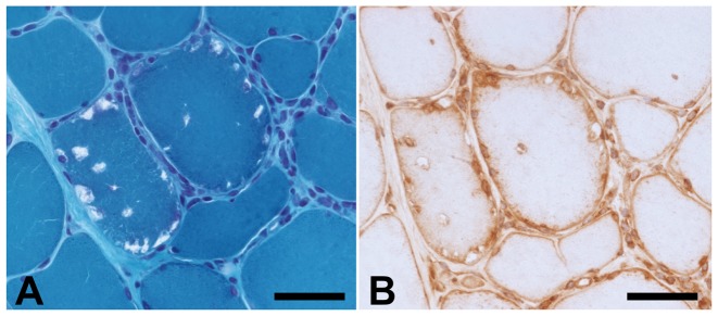
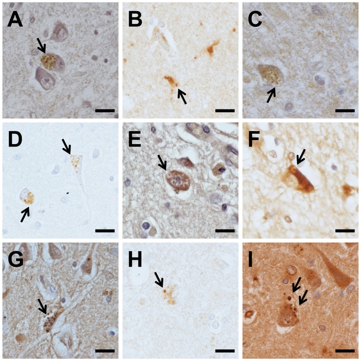
Rimmed vacuoles (RVs) are round-oval cytoplasmic inclusions, detected in muscle cells of patients with myopathies, such as inclusion body myositis (IBM) and distal myopathy with RVs (DMRV). Granulovacuolar degeneration (GVD) bodies are spherical vacuoles containing argentophilic and hematoxyphilic granules, and are one of the pathological hallmarks commonly found in hippocampal pyramidal neurons of patients with aging-related neurodegenerative diseases, such as Alzheimer's disease and Parkinson's disease. These diseases are common in the elderly and share some pathological features. Therefore, we hypothesized that mechanisms of vacuolar formation in RVs and GVD bodies are common despite their role in two differing pathologies. We explored the components of RVs by immunohistochemistry, using antibodies for GVD markers.
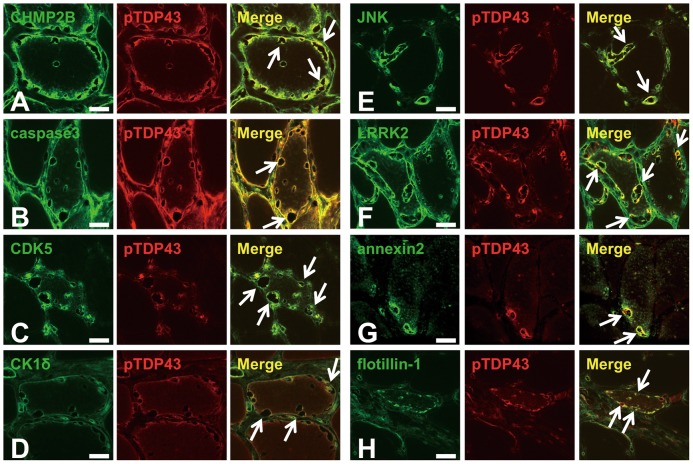
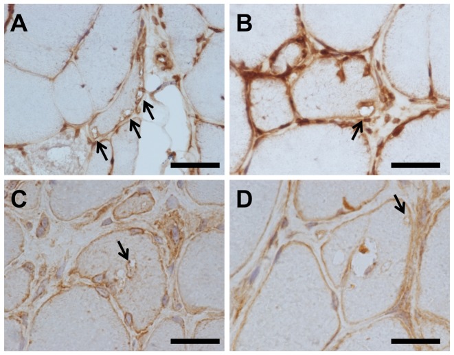
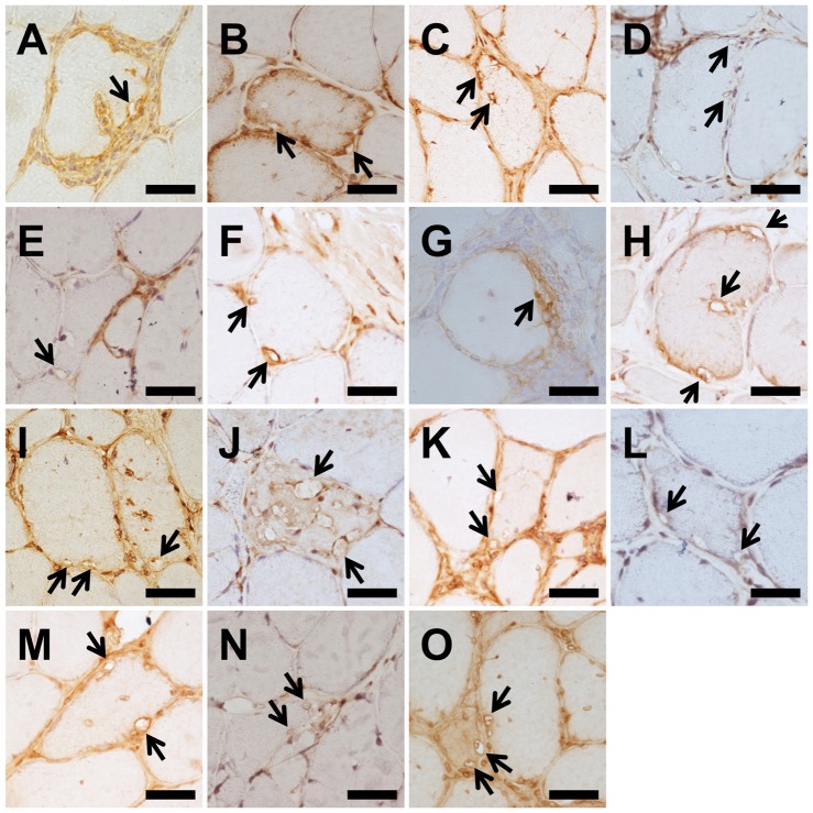
Subjects included one AD case, eight cases of sporadic IBM, and three cases of DMRV. We compared immunoreactivity and staining patterns for GVD markers. These markers included: (1) tau-modifying proteins (caspase 3, cyclin-dependent kinase 5 [CDK5], casein kinase 1δ [CK1δ], and c-jun N-terminal kinase [JNK]), (2) lipid raft-associated materials (annexin 2, leucine-rich repeat kinase 2 [LRRK2], and flotillin-1), and (3) other markers (charged multi-vesicular body protein 2B [CHMP2B] and phosphorylated transactive response DNA binding protein-43 [pTDP43]) in both GVD bodies and RVs. Furthermore, we performed double staining of each GVD marker with pTDP43 to verify the co-localization.
GVD markers, including lipid raft-associated proteins and tau kinases, were detected in RVs. CHMP2B, pTDP43, caspase 3, LRRK2, annexin 2 and flotillin-1 were detected on the rim and were diffusely distributed in the cytoplasm of RV-positive fibers. CDK5, CK1δ and JNK were detected only on the rim. In double staining experiments, all GVD markers colocalized with pTDP43 in RVs.
These results suggest that RVs of muscle cells and GVD bodies of neurons share a number of molecules, such as raft-related proteins and tau-modifying proteins.
Rimmed 空泡(RVs)是圆形或椭圆形的细胞质内含物,在肌病患者的肌肉细胞中检测到,如包涵体肌炎(IBM)和伴有 RVs 的远端肌病(DMRV)。颗粒空泡变性(GVD)体是含有银染和嗜碱性颗粒的球形空泡,是与衰老相关的神经退行性疾病(如阿尔茨海默病和帕金森病)患者海马锥体神经元中常见的病理标志物之一。这些疾病在老年人中很常见,并且具有一些共同的病理特征。因此,我们假设尽管 RVs 和 GVD 体在两种不同的病理中发挥作用,但它们的空泡形成机制是相同的。我们通过免疫组织化学方法使用 GVD 标志物抗体来探索 RVs 的成分。
研究对象包括 1 例 AD 病例、8 例散发性 IBM 病例和 3 例 DMRV 病例。我们比较了 GVD 标志物的免疫反应性和染色模式。这些标志物包括:(1)tau 修饰蛋白(半胱天冬酶 3、细胞周期蛋白依赖性激酶 5 [CDK5]、酪蛋白激酶 1δ [CK1δ] 和 c-jun N 末端激酶 [JNK])、(2)脂筏相关物质(膜联蛋白 2、富含亮氨酸重复激酶 2 [LRRK2] 和浮球素-1)和(3)其他标志物(带电多泡体蛋白 2B [CHMP2B] 和磷酸化转录激活反应 DNA 结合蛋白 43 [pTDP43])在 GVD 体和 RVs 中。此外,我们对每个 GVD 标志物与 pTDP43 进行双重染色,以验证共定位。
在 RVs 中检测到 GVD 标志物,包括脂筏相关蛋白和 tau 激酶。CHMP2B、pTDP43、半胱天冬酶 3、LRRK2、膜联蛋白 2 和浮球素-1 在边缘检测到,并在 RV 阳性纤维的细胞质中弥散分布。CDK5、CK1δ 和 JNK 仅在边缘检测到。在双重染色实验中,所有 GVD 标志物均与 RVs 中的 pTDP43 共定位。
这些结果表明,肌细胞的 RV 和神经元的 GVD 体共享许多分子,如筏相关蛋白和 tau 修饰蛋白。