• 文献检索
  • 文档翻译
  • 深度研究
  • 学术资讯
  • Suppr Zotero 插件Zotero 插件
  • 邀请有礼
  • 套餐&价格
  • 历史记录
应用&插件
Suppr Zotero 插件Zotero 插件浏览器插件Mac 客户端Windows 客户端微信小程序
定价
高级版会员购买积分包购买API积分包
服务
文献检索文档翻译深度研究API 文档MCP 服务
关于我们
关于 Suppr公司介绍联系我们用户协议隐私条款
关注我们

Suppr 超能文献

核心技术专利:CN118964589B侵权必究
粤ICP备2023148730 号-1Suppr @ 2026

文献检索

告别复杂PubMed语法,用中文像聊天一样搜索,搜遍4000万医学文献。AI智能推荐,让科研检索更轻松。

立即免费搜索

文件翻译

保留排版,准确专业,支持PDF/Word/PPT等文件格式,支持 12+语言互译。

免费翻译文档

深度研究

AI帮你快速写综述,25分钟生成高质量综述,智能提取关键信息,辅助科研写作。

立即免费体验

朊病毒感染的器官型鼠小脑培养物中浦肯野细胞树突棘密度的早期增加和晚期减少。

Early increase and late decrease of purkinje cell dendritic spine density in prion-infected organotypic mouse cerebellar cultures.

机构信息

Centre for Prions and Protein Folding Diseases, University of Alberta, Edmonton, Canada.

出版信息

PLoS One. 2013 Dec 2;8(12):e81776. doi: 10.1371/journal.pone.0081776. eCollection 2013.

DOI:10.1371/journal.pone.0081776
PMID:24312586
原文链接:https://pmc.ncbi.nlm.nih.gov/articles/PMC3847088/
Abstract

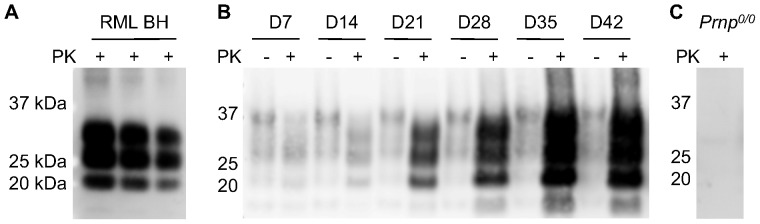
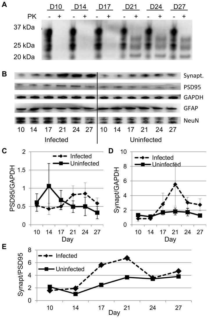
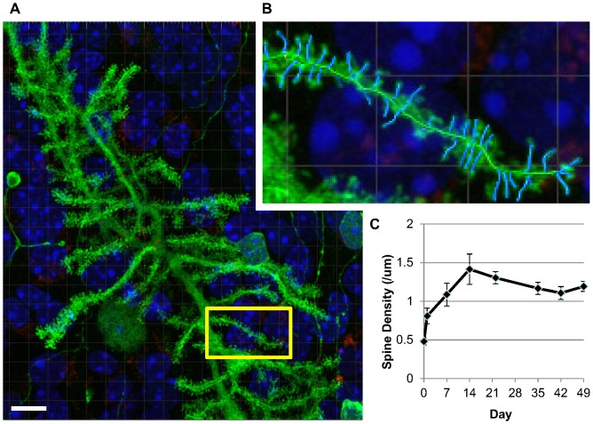
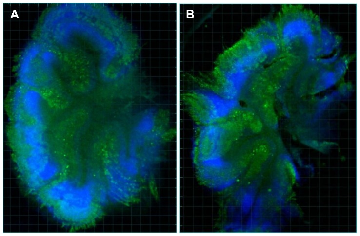
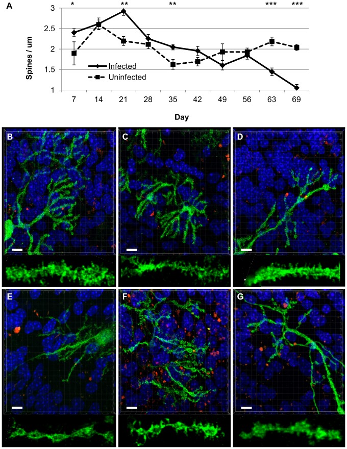
Prion diseases are infectious neurodegenerative diseases associated with the accumulation of protease-resistant prion protein, neuronal loss, spongiform change and astrogliosis. In the mouse model, the loss of dendritic spines is one of the earliest pathological changes observed in vivo, occurring 4-5 weeks after the first detection of protease-resistant prion protein in the brain. While there are cell culture models of prion infection, most do not recapitulate the neuropathology seen in vivo. Only the recently developed prion organotypic slice culture assay has been reported to undergo neuronal loss and the development of some aspects of prion pathology, namely small vacuolar degeneration and tubulovesicular bodies. Given the rapid replication of prions in this system, with protease-resistant prion protein detectable by 21 days, we investigated whether the dendritic spine loss and altered dendritic morphology seen in prion disease might also develop within the lifetime of this culture system. Indeed, six weeks after first detection of protease-resistant prion protein in tga20 mouse cerebellar slice cultures infected with RML prion strain, we found a statistically significant loss of Purkinje cell dendritic spines and altered dendritic morphology in infected cultures, analogous to that seen in vivo. In addition, we found a transient but statistically significant increase in Purkinje cell dendritic spine density during infection, at the time when protease-resistant prion protein was first detectable in culture. Our findings support the use of this slice culture system as one which recapitulates prion disease pathology and one which may facilitate study of the earliest stages of prion disease pathogenesis.

摘要

朊病毒病是一种传染性神经退行性疾病,与蛋白酶抗性朊病毒蛋白的积累、神经元丧失、海绵状改变和星形胶质细胞增生有关。在小鼠模型中,树突棘的丧失是体内最早观察到的病理变化之一,发生在大脑中首次检测到蛋白酶抗性朊病毒蛋白后的 4-5 周。虽然有朊病毒感染的细胞培养模型,但大多数模型不能重现体内观察到的神经病理学变化。只有最近开发的朊病毒器官型切片培养测定法被报道会发生神经元丧失和朊病毒病理学的某些方面的发展,即小空泡变性和管状囊泡体。鉴于该系统中朊病毒的快速复制,在 21 天内可检测到蛋白酶抗性朊病毒蛋白,我们研究了朊病毒病中所见的树突棘丧失和树突形态改变是否也可能在该培养系统的寿命内发展。事实上,在 tga20 小鼠小脑切片培养物中感染 RML 朊病毒株后,首次检测到蛋白酶抗性朊病毒蛋白的 6 周后,我们发现感染培养物中的浦肯野细胞树突棘显著丧失,树突形态改变,与体内所见相似。此外,我们发现感染期间浦肯野细胞树突棘密度短暂但具有统计学意义的增加,此时在培养物中首次检测到蛋白酶抗性朊病毒蛋白。我们的研究结果支持使用该切片培养系统来重现朊病毒病病理学,并可能有助于研究朊病毒病发病机制的早期阶段。

https://cdn.ncbi.nlm.nih.gov/pmc/blobs/1898/3847088/85a11c6f1a3a/pone.0081776.g005.jpg
https://cdn.ncbi.nlm.nih.gov/pmc/blobs/1898/3847088/7b26c70eb121/pone.0081776.g001.jpg
https://cdn.ncbi.nlm.nih.gov/pmc/blobs/1898/3847088/874fd14339f5/pone.0081776.g002.jpg
https://cdn.ncbi.nlm.nih.gov/pmc/blobs/1898/3847088/8138823c27ec/pone.0081776.g003.jpg
https://cdn.ncbi.nlm.nih.gov/pmc/blobs/1898/3847088/a18e109ce89e/pone.0081776.g004.jpg
https://cdn.ncbi.nlm.nih.gov/pmc/blobs/1898/3847088/85a11c6f1a3a/pone.0081776.g005.jpg

相似文献

1
Early increase and late decrease of purkinje cell dendritic spine density in prion-infected organotypic mouse cerebellar cultures.朊病毒感染的器官型鼠小脑培养物中浦肯野细胞树突棘密度的早期增加和晚期减少。
PLoS One. 2013 Dec 2;8(12):e81776. doi: 10.1371/journal.pone.0081776. eCollection 2013.
2
Serial changes in granuloprival cerebellar cultures after transplantation with granule cells and glia: a timed ultrastructural study.颗粒细胞和胶质细胞移植后颗粒细胞缺乏型小脑培养物的系列变化:一项定时超微结构研究。
Neuroscience. 1997 Apr;77(3):695-711. doi: 10.1016/s0306-4522(96)00546-5.
3
A Neuronal Culture System to Detect Prion Synaptotoxicity.一种用于检测朊病毒突触毒性的神经元培养系统。
PLoS Pathog. 2016 May 26;12(5):e1005623. doi: 10.1371/journal.ppat.1005623. eCollection 2016 May.
4
The prion organotypic slice culture assay--POSCA.朊病毒器官型切片培养检测法——POSCA
Nat Protoc. 2008;3(4):555-62. doi: 10.1038/nprot.2008.13.
5
Prions activate a p38 MAPK synaptotoxic signaling pathway.朊病毒激活 p38 MAPK 突触毒性信号通路。
PLoS Pathog. 2018 Sep 20;14(9):e1007283. doi: 10.1371/journal.ppat.1007283. eCollection 2018 Sep.
6
Corticotropin-releasing factor and urocortin regulate spine and synapse formation: structural basis for stress-induced neuronal remodeling and pathology.促肾上腺皮质素释放因子和尿皮质素调节脊柱和突触形成:应激诱导的神经元重塑和病理学的结构基础。
Mol Psychiatry. 2013 Jan;18(1):86-92. doi: 10.1038/mp.2012.43. Epub 2012 May 1.
7
Cerebellar culture models of dendritic spine proliferation after transplantation of glia.胶质细胞移植后树突棘增殖的小脑培养模型。
J Neural Transplant Plast. 1997 Jan-Mar;6(1):1-10. doi: 10.1155/NP.1997.1.
8
Reorganization of organotypic cultures of mouse cerebellum exposed to cytosine arabinoside: a timed ultrastructural study.暴露于阿糖胞苷的小鼠小脑器官型培养物的重组:一项定时超微结构研究。
J Comp Neurol. 1991 Nov 8;313(2):193-212. doi: 10.1002/cne.903130202.
9
Deposition pattern and subcellular distribution of disease-associated prion protein in cerebellar organotypic slice cultures infected with scrapie.感染羊瘙痒病的小脑器官型切片培养物中疾病相关朊病毒蛋白的沉积模式和亚细胞分布
Front Neurosci. 2015 Nov 4;9:410. doi: 10.3389/fnins.2015.00410. eCollection 2015.
10
Dendritic pathology in prion disease starts at the synaptic spine.朊病毒病中的树突状病理始于突触棘。
J Neurosci. 2007 Jun 6;27(23):6224-33. doi: 10.1523/JNEUROSCI.5062-06.2007.

引用本文的文献

1
Prion replication in organotypic brain slice cultures is distinct from in vivo inoculation and is species dependent.朊病毒在器官型脑片培养物中的复制不同于体内接种,且具有种属依赖性。
Acta Neuropathol Commun. 2025 Apr 30;13(1):86. doi: 10.1186/s40478-025-01999-w.
2
Distinct patterns of prion strain deposition and toxicity in a novel whole brain organotypic slice culture system.新型全脑器官型切片培养系统中朊病毒株沉积和毒性的独特模式。
Sci Rep. 2025 Feb 8;15(1):4681. doi: 10.1038/s41598-025-88861-0.
3
Single-cell transcriptomics unveils molecular signatures of neuronal vulnerability in a mouse model of prion disease that overlap with Alzheimer's disease.

本文引用的文献

1
Development of the cerebellum: from gene expression patterns to circuit maps.小脑的发育:从基因表达模式到神经回路图谱
Wiley Interdiscip Rev Dev Biol. 2013 Jan-Feb;2(1):149-64. doi: 10.1002/wdev.65. Epub 2012 May 7.
2
Brain region specific pre-synaptic and post-synaptic degeneration are early components of neuropathology in prion disease.脑区特异的突触前和突触后变性是朊病毒病神经病理学的早期组成部分。
PLoS One. 2013;8(1):e55004. doi: 10.1371/journal.pone.0055004. Epub 2013 Jan 30.
3
Prion pathogenesis is faithfully reproduced in cerebellar organotypic slice cultures.
单细胞转录组学揭示了朊病毒病小鼠模型中神经元易损性的分子特征,这些特征与阿尔茨海默病重叠。
Nat Commun. 2024 Nov 23;15(1):10174. doi: 10.1038/s41467-024-54579-2.
4
Mechanisms of prion-induced toxicity.朊病毒诱导毒性的机制。
Cell Tissue Res. 2023 Apr;392(1):81-96. doi: 10.1007/s00441-022-03683-0. Epub 2022 Sep 7.
5
Neurons and Astrocytes Elicit Brain Region Specific Transcriptional Responses to Prion Disease in the Murine CA1 and Thalamus.神经元和星形胶质细胞引发小鼠CA1区和丘脑对朊病毒病的脑区特异性转录反应。
Front Neurosci. 2022 May 16;16:918811. doi: 10.3389/fnins.2022.918811. eCollection 2022.
6
Genetically engineered cellular models of prion propagation.朊病毒传播的基因工程细胞模型。
Cell Tissue Res. 2023 Apr;392(1):63-80. doi: 10.1007/s00441-022-03630-z. Epub 2022 May 18.
7
Reflections on Cerebellar Neuropathology in Classical Scrapie.经典瘙痒病小脑神经病理学的反思。
Biomolecules. 2021 Apr 28;11(5):649. doi: 10.3390/biom11050649.
8
From Cell Culture to Organoids-Model Systems for Investigating Prion Strain Characteristics.从细胞培养到类器官——用于研究朊病毒株特性的模型系统
Biomolecules. 2021 Jan 14;11(1):106. doi: 10.3390/biom11010106.
9
POSCAbilities: The Application of the Prion Organotypic Slice Culture Assay to Neurodegenerative Disease Research.POSCAbilities:Prion 器官型切片培养测定在神经退行性疾病研究中的应用。
Biomolecules. 2020 Jul 20;10(7):1079. doi: 10.3390/biom10071079.
10
Mutant prion proteins increase calcium permeability of AMPA receptors, exacerbating excitotoxicity.突变朊病毒蛋白增加 AMPA 受体的钙通透性,从而加剧兴奋性毒性。
PLoS Pathog. 2020 Jul 16;16(7):e1008654. doi: 10.1371/journal.ppat.1008654. eCollection 2020 Jul.
朊病毒病的发病机制在小脑器官型切片培养中得到了准确再现。
PLoS Pathog. 2012;8(11):e1002985. doi: 10.1371/journal.ppat.1002985. Epub 2012 Nov 1.
4
Sustained translational repression by eIF2α-P mediates prion neurodegeneration.eIF2α-P 介导的持续翻译抑制导致朊病毒神经退行性变。
Nature. 2012 May 6;485(7399):507-11. doi: 10.1038/nature11058.
5
Structural dynamics of dendritic spines in memory and cognition.树突棘的结构动力学在记忆和认知中的作用。
Trends Neurosci. 2010 Mar;33(3):121-9. doi: 10.1016/j.tins.2010.01.001.
6
Strain-associated variations in abnormal PrP trafficking of sheep scrapie.绵羊瘙痒病异常朊蛋白转运中与毒株相关的变异
Brain Pathol. 2009 Jan;19(1):1-11. doi: 10.1111/j.1750-3639.2008.00150.x. Epub 2008 Apr 8.
7
The prion organotypic slice culture assay--POSCA.朊病毒器官型切片培养检测法——POSCA
Nat Protoc. 2008;3(4):555-62. doi: 10.1038/nprot.2008.13.
8
A versatile prion replication assay in organotypic brain slices.一种用于器官型脑片的通用朊病毒复制检测方法。
Nat Neurosci. 2008 Jan;11(1):109-17. doi: 10.1038/nn2028. Epub 2007 Dec 9.
9
Dendritic pathology in prion disease starts at the synaptic spine.朊病毒病中的树突状病理始于突触棘。
J Neurosci. 2007 Jun 6;27(23):6224-33. doi: 10.1523/JNEUROSCI.5062-06.2007.
10
Formation of native prions from minimal components in vitro.在体外由最小成分形成天然朊病毒。
Proc Natl Acad Sci U S A. 2007 Jun 5;104(23):9741-6. doi: 10.1073/pnas.0702662104. Epub 2007 May 29.