• 文献检索
  • 文档翻译
  • 深度研究
  • 学术资讯
  • Suppr Zotero 插件Zotero 插件
  • 邀请有礼
  • 套餐&价格
  • 历史记录
应用&插件
Suppr Zotero 插件Zotero 插件浏览器插件Mac 客户端Windows 客户端微信小程序
定价
高级版会员购买积分包购买API积分包
服务
文献检索文档翻译深度研究API 文档MCP 服务
关于我们
关于 Suppr公司介绍联系我们用户协议隐私条款
关注我们

Suppr 超能文献

核心技术专利:CN118964589B侵权必究
粤ICP备2023148730 号-1Suppr @ 2026

文献检索

告别复杂PubMed语法,用中文像聊天一样搜索,搜遍4000万医学文献。AI智能推荐,让科研检索更轻松。

立即免费搜索

文件翻译

保留排版,准确专业,支持PDF/Word/PPT等文件格式,支持 12+语言互译。

免费翻译文档

深度研究

AI帮你快速写综述,25分钟生成高质量综述,智能提取关键信息,辅助科研写作。

立即免费体验

内肝微循环障碍、实质缺氧和 NOX4 上调导致脂多糖和 D-半乳糖胺诱导的大鼠急性肝衰竭后肝细胞凋亡的区域性差异。

Intrahepatic microcirculatory disorder, parenchymal hypoxia and NOX4 upregulation result in zonal differences in hepatocyte apoptosis following lipopolysaccharide- and D-galactosamine-induced acute liver failure in rats.

机构信息

Department of Medicine and Bioregulatory Science, Graduate School of Medical Sciences, Kyushu University, Fukuoka 812‑8582, Japan.

Faculty of Pharmaceutical Sciences, Fukuoka University, Fukuoka 814-0180, Japan.

出版信息

Int J Mol Med. 2014 Feb;33(2):254-62. doi: 10.3892/ijmm.2013.1573. Epub 2013 Dec 3.

DOI:10.3892/ijmm.2013.1573
PMID:24317376
原文链接:https://pmc.ncbi.nlm.nih.gov/articles/PMC3896462/
Abstract

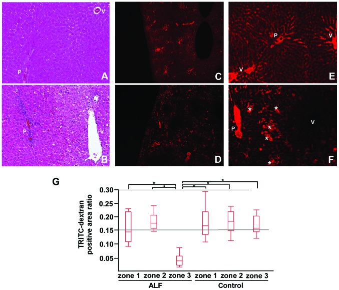
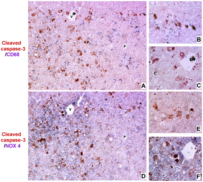
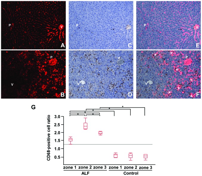
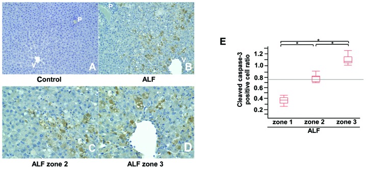
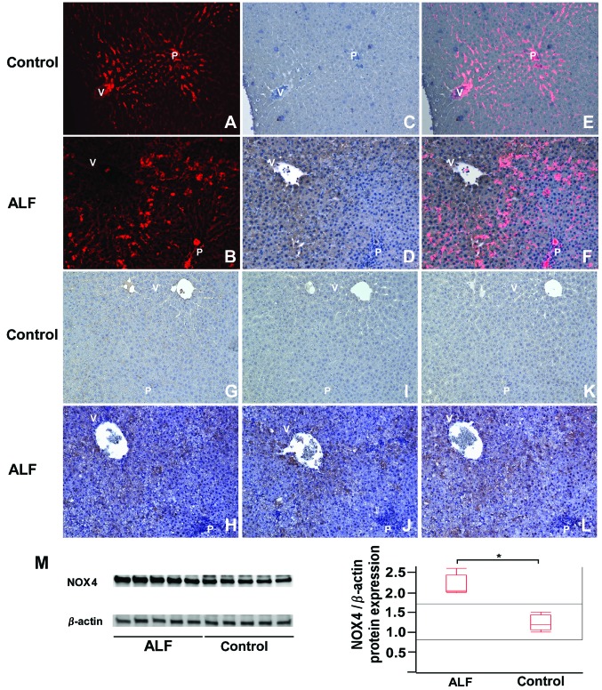
Although the mechanisms responsible for acute liver failure (ALF) have not yet been fully elucidated, studies have indicated that intrahepatic macrophage activation plays an important role in the pathogenesis of ALF through intrahepatic microcirculatory disorder and consequent parenchymal cell death. Intrahepatic microcirculatory disorder has been demonstrated in animal models using intravital microscopy; however, the limitations of this method include simultaneously evaluating blood flow and the surrounding pathological changes. Therefore, in this study, we devised a novel method involving tetramethylrhodamine isothiocyanate (TRITC)-dextran administration for the pathological assessment of hepatic microcirculation. In addition, we aimed to elucidate the mechanisms through which intrahepatic microcirculatory disorder progresses with relation to activated macrophages. ALF was induced in Wistar rats by exposure to lipopolysaccharide and D-galactosamine. Intrahepatic microcirculation and microcirculatory disorder in zone 3 (pericentral zone) of the livers of rats with ALF was observed. Immunohistochemical examinations in conjunction with TRITC-dextran images revealed that the macrophages were mainly distributed in zone 2 (intermediate zone), while cleaved caspase-3-positive hepatocytes, pimonidazole and hypoxia-inducible factor 1-α were abundant in zone 3. We also found that 4-hydroxy-2-nonenal and nicotinamide adenine dinucleotide phosphate oxidase (NOX)4-positive cells were predominantly located in the zone 3 parenchyma. The majority of apoptotic hepatocytes in zone 3 were co-localized with NOX4. Our results revealed that the apoptotic cells in zone 3 were a result of hypoxic conditions induced by intrahepatic microcirculatory disorder, and were not induced by activated macrophages. The increased levels of oxidative stress in zone 3 may contribute to the progression of hepatocyte apoptosis.

摘要

虽然导致急性肝衰竭(ALF)的确切机制尚未完全阐明,但研究表明,肝内巨噬细胞的激活通过肝内微循环障碍和随后的实质细胞死亡在 ALF 的发病机制中发挥重要作用。在动物模型中,已经使用活体显微镜证实了肝内微循环障碍;然而,这种方法的局限性在于同时评估血流和周围的病理变化。因此,在这项研究中,我们设计了一种新的方法,涉及四甲基罗丹明异硫氰酸酯(TRITC)-葡聚糖给药,用于肝微循环的病理评估。此外,我们旨在阐明与激活的巨噬细胞有关的肝内微循环障碍进展的机制。通过暴露于脂多糖和 D-半乳糖胺诱导 Wistar 大鼠发生 ALF。观察到 ALF 大鼠肝脏 3 区(中心区周围区)的肝内微循环和微循环障碍。与 TRITC-葡聚糖图像结合的免疫组织化学检查显示,巨噬细胞主要分布在 2 区(中间区),而 cleaved caspase-3 阳性肝细胞、pimonidazole 和缺氧诱导因子 1-α在 3 区丰富。我们还发现 4-羟基-2-壬烯醛和烟酰胺腺嘌呤二核苷酸磷酸氧化酶(NOX)4 阳性细胞主要位于 3 区实质。3 区凋亡的肝细胞大多数与 NOX4 共定位。我们的结果表明,3 区的凋亡细胞是由肝内微循环障碍引起的缺氧条件导致的,而不是由激活的巨噬细胞引起的。3 区氧化应激水平的升高可能导致肝细胞凋亡的进展。

https://cdn.ncbi.nlm.nih.gov/pmc/blobs/c491/3896462/152eb14c6700/IJMM-33-02-0254-g04.jpg
https://cdn.ncbi.nlm.nih.gov/pmc/blobs/c491/3896462/da3cec00b96c/IJMM-33-02-0254-g00.jpg
https://cdn.ncbi.nlm.nih.gov/pmc/blobs/c491/3896462/22f7e2394f91/IJMM-33-02-0254-g01.jpg
https://cdn.ncbi.nlm.nih.gov/pmc/blobs/c491/3896462/e3e33ebf3b0a/IJMM-33-02-0254-g02.jpg
https://cdn.ncbi.nlm.nih.gov/pmc/blobs/c491/3896462/fd22072d22dd/IJMM-33-02-0254-g03.jpg
https://cdn.ncbi.nlm.nih.gov/pmc/blobs/c491/3896462/152eb14c6700/IJMM-33-02-0254-g04.jpg

相似文献

1
Intrahepatic microcirculatory disorder, parenchymal hypoxia and NOX4 upregulation result in zonal differences in hepatocyte apoptosis following lipopolysaccharide- and D-galactosamine-induced acute liver failure in rats.内肝微循环障碍、实质缺氧和 NOX4 上调导致脂多糖和 D-半乳糖胺诱导的大鼠急性肝衰竭后肝细胞凋亡的区域性差异。
Int J Mol Med. 2014 Feb;33(2):254-62. doi: 10.3892/ijmm.2013.1573. Epub 2013 Dec 3.
2
Therapeutic benefits of apocynin in mice with lipopolysaccharide/D-galactosamine-induced acute liver injury via suppression of the late stage pro-apoptotic AMPK/JNK pathway.野靛碱通过抑制晚期促凋亡的 AMPK/JNK 通路对脂多糖/半乳糖胺诱导的急性肝损伤小鼠的治疗作用。
Biomed Pharmacother. 2020 May;125:110020. doi: 10.1016/j.biopha.2020.110020. Epub 2020 Feb 25.
3
Inhibitory effect of oxymatrine on hepatocyte apoptosis TLR4/PI3K/Akt/GSK-3β signaling pathway.氧化苦参碱对肝细胞凋亡TLR4/PI3K/Akt/GSK-3β信号通路的抑制作用。
World J Gastroenterol. 2017 Jun 7;23(21):3839-3849. doi: 10.3748/wjg.v23.i21.3839.
4
Cystathionine γ-lyase deficiency protects mice from galactosamine/lipopolysaccharide-induced acute liver failure.胱硫醚γ-裂解酶缺乏可保护小鼠免于半乳糖胺/脂多糖诱导的急性肝衰竭。
Antioxid Redox Signal. 2014 Jan 10;20(2):204-16. doi: 10.1089/ars.2013.5354. Epub 2013 Aug 22.
5
Promotion of mitochondrial energy metabolism during hepatocyte apoptosis in a rat model of acute liver failure.急性肝衰竭大鼠模型中肝细胞凋亡期间线粒体能量代谢的促进作用
Mol Med Rep. 2015 Oct;12(4):5035-41. doi: 10.3892/mmr.2015.4029. Epub 2015 Jul 2.
6
Peroxisome proliferator-activated receptor alpha acts as a mediator of endoplasmic reticulum stress-induced hepatocyte apoptosis in acute liver failure.过氧化物酶体增殖物激活受体α作为内质网应激诱导急性肝衰竭中肝细胞凋亡的介质。
Dis Model Mech. 2016 Jul 1;9(7):799-809. doi: 10.1242/dmm.023242. Epub 2016 May 26.
7
Protective Role of 4-Octyl Itaconate in Murine LPS/D-GalN-Induced Acute Liver Failure via Inhibiting Inflammation, Oxidative Stress, and Apoptosis.4-辛基衣康酸酯通过抑制炎症、氧化应激和细胞凋亡对小鼠脂多糖/半乳糖胺诱导的急性肝衰竭的保护作用。
Oxid Med Cell Longev. 2021 Aug 17;2021:9932099. doi: 10.1155/2021/9932099. eCollection 2021.
8
Endoplasmic reticulum stress-activated glycogen synthase kinase 3β aggravates liver inflammation and hepatotoxicity in mice with acute liver failure.内质网应激激活的糖原合酶激酶 3β加剧急性肝衰竭小鼠的肝脏炎症和肝毒性。
Inflammation. 2015;38(3):1151-65. doi: 10.1007/s10753-014-0080-2.
9
[Effects of granulocyte colony-stimulating factor on hepatocyte apoptosis in acute liver failure: experiment with rats].[粒细胞集落刺激因子对急性肝衰竭大鼠肝细胞凋亡的影响:实验研究]
Zhonghua Yi Xue Za Zhi. 2008 Apr 8;88(14):980-4.
10
Salvage effect of E5564, Toll-like receptor 4 antagonist on d-galactosamine and lipopolysaccharide-induced acute liver failure in rats.E5564,Toll 样受体 4 拮抗剂对 D-半乳糖胺和脂多糖诱导的大鼠急性肝衰竭的挽救作用。
J Gastroenterol Hepatol. 2010 May;25(5):1009-12. doi: 10.1111/j.1440-1746.2009.06145.x.

引用本文的文献

1
Cardiac Hepatopathy: New Perspectives on Old Problems through a Prism of Endogenous Metabolic Regulations by Hepatokines.心性肝病:通过肝细胞因子内源性代谢调节视角对老问题的新见解。
Antioxidants (Basel). 2023 Feb 17;12(2):516. doi: 10.3390/antiox12020516.
2
Effect of Melatonin on the Expression of LYVE-1 Receptor in Liver Sinusoidal Endothelial Cells of Mice with the Light-Induced Functional Pinealectomy Model.褪黑素对光诱导功能性松果体切除模型小鼠肝脏窦内皮细胞 LYVE-1 受体表达的影响。
Bull Exp Biol Med. 2022 Jun;173(2):182-187. doi: 10.1007/s10517-022-05515-1. Epub 2022 Jun 23.
3
High-resolution three-dimensional visualization of hepatic sinusoids in cirrhotic rats via serial histological sections.

本文引用的文献

1
Etiology and prognosis of fulminant hepatitis and late-onset hepatic failure in Japan: Summary of the annual nationwide survey between 2004 and 2009.日本暴发性肝炎和迟发性肝衰竭的病因和预后:2004 年至 2009 年全国年度调查总结。
Hepatol Res. 2013 Feb;43(2):97-105. doi: 10.1111/j.1872-034X.2012.01105.x.
2
NADPH oxidase 4 mediates insulin-stimulated HIF-1α and VEGF expression, and angiogenesis in vitro.烟酰胺腺嘌呤二核苷酸磷酸氧化酶4介导胰岛素刺激的低氧诱导因子-1α和血管内皮生长因子表达以及体外血管生成。
PLoS One. 2012;7(10):e48393. doi: 10.1371/journal.pone.0048393. Epub 2012 Oct 29.
3
From form to function: the role of Nox4 in the cardiovascular system.
通过连续组织切片实现肝硬化大鼠肝窦的高分辨率三维可视化。
Histol Histopathol. 2021 May;36(5):577-586. doi: 10.14670/HH-18-339. Epub 2021 Apr 14.
4
Switching from Fatty Acid Oxidation to Glycolysis Improves the Outcome of Acute-On-Chronic Liver Failure.从脂肪酸氧化转变为糖酵解可改善慢加急性肝衰竭的预后。
Adv Sci (Weinh). 2020 Feb 13;7(7):1902996. doi: 10.1002/advs.201902996. eCollection 2020 Apr.
5
Triptriolide Alleviates Lipopolysaccharide-Induced Liver Injury by Nrf2 and NF-κB Signaling Pathways.雷公藤甲素通过Nrf2和NF-κB信号通路减轻脂多糖诱导的肝损伤。
Front Pharmacol. 2018 Aug 29;9:999. doi: 10.3389/fphar.2018.00999. eCollection 2018.
6
Does Hypoxia Cause Carcinogenic Iron Accumulation in Alcoholic Liver Disease (ALD)?缺氧会导致酒精性肝病(ALD)中致癌性铁蓄积吗?
Cancers (Basel). 2017 Oct 25;9(11):145. doi: 10.3390/cancers9110145.
7
Pharmacological inhibition of NOX4 ameliorates alcohol-induced liver injury in mice through improving oxidative stress and mitochondrial function.药理学抑制 NOX4 可通过改善氧化应激和线粒体功能来减轻小鼠的酒精性肝损伤。
Biochim Biophys Acta Gen Subj. 2017 Jan;1861(1 Pt A):2912-2921. doi: 10.1016/j.bbagen.2016.09.009. Epub 2016 Sep 12.
8
Effect of silibinin and vitamin E on the ASK1-p38 MAPK pathway in D-galactosamine/lipopolysaccharide induced hepatotoxicity.水飞蓟宾和维生素E对D-半乳糖胺/脂多糖诱导的肝毒性中ASK1-p38丝裂原活化蛋白激酶通路的影响。
Exp Biol Med (Maywood). 2016 Jun;241(11):1250-7. doi: 10.1177/1535370216636719. Epub 2016 Mar 3.
9
Hepatic Shock Differential Diagnosis and Risk Factors: A Review Article.肝性休克的鉴别诊断与危险因素:一篇综述文章
Hepat Mon. 2015 Oct 10;15(10):e27063. doi: 10.5812/hepatmon.27063. eCollection 2015 Oct.
从形态到功能:Nox4 在心血管系统中的作用。
Front Physiol. 2012 Nov 1;3:412. doi: 10.3389/fphys.2012.00412. eCollection 2012.
4
NADPH oxidase NOX4 mediates stellate cell activation and hepatocyte cell death during liver fibrosis development.NADPH 氧化酶 NOX4 在肝纤维化发展过程中介导星状细胞激活和肝细胞死亡。
PLoS One. 2012;7(9):e45285. doi: 10.1371/journal.pone.0045285. Epub 2012 Sep 26.
5
Development of an accurate index for predicting outcomes of patients with acute liver failure.开发一种准确的指数,用于预测急性肝衰竭患者的预后。
Gastroenterology. 2012 Nov;143(5):1237-1243. doi: 10.1053/j.gastro.2012.07.113. Epub 2012 Aug 8.
6
NADPH oxidase inhibitors: a decade of discovery from Nox2ds to HTS.NADPH 氧化酶抑制剂:从 Nox2ds 到高通量筛选的十年发现。
Cell Mol Life Sci. 2012 Jul;69(14):2315-25. doi: 10.1007/s00018-012-1009-2. Epub 2012 May 15.
7
Antithrombin III injection via the portal vein suppresses liver damage.经门静脉注射抗凝血酶 III 可抑制肝损伤。
World J Gastroenterol. 2012 Apr 28;18(16):1884-91. doi: 10.3748/wjg.v18.i16.1884.
8
NOX isoforms and reactive oxygen species in vascular health.血管健康中的NADPH氧化酶亚型与活性氧物质
Mol Interv. 2011 Feb;11(1):27-35. doi: 10.1124/mi.11.1.5.
9
Acetaminophen hepatotoxicity and HIF-1α induction in acetaminophen toxicity in mice occurs without hypoxia.对乙酰氨基酚诱导的肝毒性和 HIF-1α 在小鼠对乙酰氨基酚中毒中的诱导作用发生在非缺氧条件下。
Toxicol Appl Pharmacol. 2011 May 1;252(3):211-20. doi: 10.1016/j.taap.2011.02.005. Epub 2011 Feb 24.
10
Myofibroblast differentiation during fibrosis: role of NAD(P)H oxidases.成纤维细胞向肌成纤维细胞分化在纤维化过程中的作用:NAD(P)H 氧化酶的作用。
Kidney Int. 2011 May;79(9):944-56. doi: 10.1038/ki.2010.516. Epub 2011 Feb 9.