• 文献检索
  • 文档翻译
  • 深度研究
  • 学术资讯
  • Suppr Zotero 插件Zotero 插件
  • 邀请有礼
  • 套餐&价格
  • 历史记录
应用&插件
Suppr Zotero 插件Zotero 插件浏览器插件Mac 客户端Windows 客户端微信小程序
定价
高级版会员购买积分包购买API积分包
服务
文献检索文档翻译深度研究API 文档MCP 服务
关于我们
关于 Suppr公司介绍联系我们用户协议隐私条款
关注我们

Suppr 超能文献

核心技术专利:CN118964589B侵权必究
粤ICP备2023148730 号-1Suppr @ 2026

文献检索

告别复杂PubMed语法,用中文像聊天一样搜索,搜遍4000万医学文献。AI智能推荐,让科研检索更轻松。

立即免费搜索

文件翻译

保留排版,准确专业,支持PDF/Word/PPT等文件格式,支持 12+语言互译。

免费翻译文档

深度研究

AI帮你快速写综述,25分钟生成高质量综述,智能提取关键信息,辅助科研写作。

立即免费体验

低浓度冈田酸毒素可促进胃和结肠上皮细胞系增殖。

Okadaic acid toxin at sublethal dose produced cell proliferation in gastric and colon epithelial cell lines.

机构信息

Laboratory of Membrane Biochemistry, Program of Physiology and Biophysics, Faculty of Medicine, University of Chile, Independencia 1027, Correo 7, Santiago 8380000, Chile.

出版信息

Mar Drugs. 2013 Dec 2;11(12):4751-60. doi: 10.3390/md11124751.

DOI:10.3390/md11124751
PMID:24317467
原文链接:https://pmc.ncbi.nlm.nih.gov/articles/PMC3877884/
Abstract

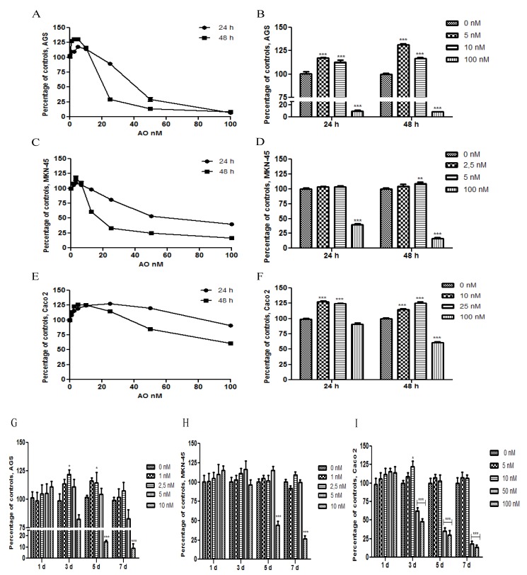
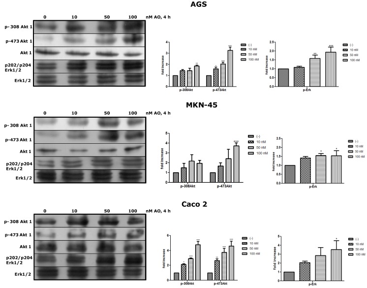
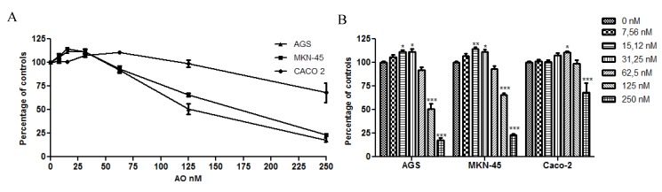
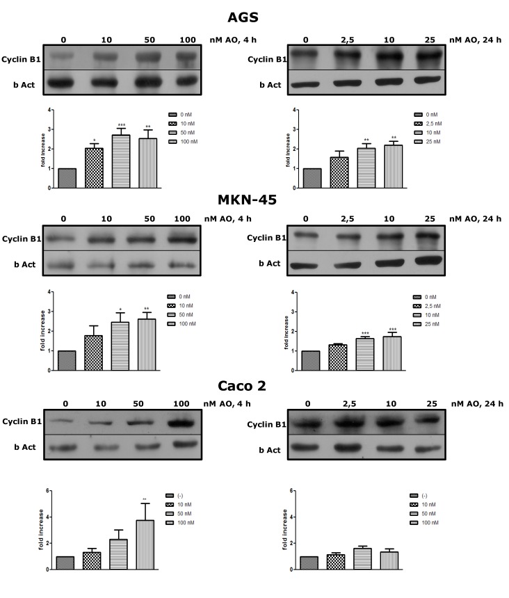
The aim of this study was to analyze the effect of Okadaic Acid (OA) on the proliferation of gastric and colon epithelial cells, the main target tissues of the toxin. We hypothesized that OA, at sublethal doses, activates multiple signaling pathways, such as Erk and Akt, through the inhibition of PP2A. To demonstrate this, we carried out curves of doses and time response against OA in AGS, MKN-45 and Caco 2 cell lines, and found an increase in the cell proliferation at sublethal doses, at 24 h or 48 h exposure. Indeed, cells can withstand high concentrations of the toxin at 4 h exposure, the time chosen considering the maximum time before total gastric emptying. We have proved that this increased proliferation is due to an overexpression of Cyclin B, a cyclin that promotes the passage from G2 to mitosis. In addition, we have demonstrated that OA induces activation of Akt and Erk in the three cells lines, showing that OA can activate pathways involved in oncogenesis. In conclusion, this study contributes to the knowledge about the possible effects of chronic OA consumption.

摘要

本研究旨在分析冈田酸(OA)对胃和结肠上皮细胞增殖的影响,这些细胞是毒素的主要靶组织。我们假设 OA 在亚致死剂量下通过抑制 PP2A 来激活多种信号通路,如 Erk 和 Akt。为了证明这一点,我们在 AGS、MKN-45 和 Caco 2 细胞系中进行了 OA 剂量和时间反应曲线,发现亚致死剂量在 24 小时或 48 小时暴露时会增加细胞增殖。事实上,细胞可以在 4 小时暴露时耐受高浓度的毒素,这个时间是在胃排空之前的最大时间考虑选择的。我们已经证明,这种增殖的增加是由于细胞周期蛋白 B 的过度表达,细胞周期蛋白 B 促进了从 G2 期到有丝分裂的过渡。此外,我们还证明了 OA 可以在这三种细胞系中诱导 Akt 和 Erk 的激活,表明 OA 可以激活参与致癌的途径。总之,本研究有助于了解慢性 OA 消费可能产生的影响。

https://cdn.ncbi.nlm.nih.gov/pmc/blobs/5d53/3877884/df7f45e20192/marinedrugs-11-04751-g004.jpg
https://cdn.ncbi.nlm.nih.gov/pmc/blobs/5d53/3877884/92c12c817225/marinedrugs-11-04751-g001.jpg
https://cdn.ncbi.nlm.nih.gov/pmc/blobs/5d53/3877884/d98a9fa78cf1/marinedrugs-11-04751-g002.jpg
https://cdn.ncbi.nlm.nih.gov/pmc/blobs/5d53/3877884/20f68cf60f32/marinedrugs-11-04751-g003.jpg
https://cdn.ncbi.nlm.nih.gov/pmc/blobs/5d53/3877884/df7f45e20192/marinedrugs-11-04751-g004.jpg

相似文献

1
Okadaic acid toxin at sublethal dose produced cell proliferation in gastric and colon epithelial cell lines.低浓度冈田酸毒素可促进胃和结肠上皮细胞系增殖。
Mar Drugs. 2013 Dec 2;11(12):4751-60. doi: 10.3390/md11124751.
2
Inhibition of PP2A, but not PP5, mediates p53 activation by low levels of okadaic acid in rat liver epithelial cells.在大鼠肝上皮细胞中,抑制蛋白磷酸酶2A(PP2A)而非蛋白磷酸酶5(PP5)介导了低水平冈田酸对p53的激活作用。
J Cell Biochem. 2006 Sep 1;99(1):241-55. doi: 10.1002/jcb.20919.
3
Genotoxicity of the marine toxin okadaic acid, in human Caco-2 cells and in mice gut cells.海洋毒素冈田酸对人结肠腺癌细胞(Caco-2细胞)和小鼠肠道细胞的遗传毒性。
Environ Toxicol. 2006 Feb;21(1):55-64. doi: 10.1002/tox.20154.
4
Danusertib, a potent pan-Aurora kinase and ABL kinase inhibitor, induces cell cycle arrest and programmed cell death and inhibits epithelial to mesenchymal transition involving the PI3K/Akt/mTOR-mediated signaling pathway in human gastric cancer AGS and NCI-N78 cells.达努塞替布是一种强效的泛极光激酶和ABL激酶抑制剂,可诱导细胞周期停滞和程序性细胞死亡,并在人胃癌AGS和NCI-N78细胞中抑制涉及PI3K/Akt/mTOR介导的信号通路的上皮-间质转化。
Drug Des Devel Ther. 2015 Mar 2;9:1293-318. doi: 10.2147/DDDT.S74964. eCollection 2015.
5
Lycopene treatment inhibits activation of Jak1/Stat3 and Wnt/β-catenin signaling and attenuates hyperproliferation in gastric epithelial cells.番茄红素处理可抑制 Jak1/Stat3 和 Wnt/β-连环蛋白信号通路的激活,并减轻胃上皮细胞的过度增殖。
Nutr Res. 2019 Oct;70:70-81. doi: 10.1016/j.nutres.2018.07.010. Epub 2018 Jul 21.
6
Okadaic acid induces Akt hyperphosphorylation and an oxidative stress-mediated cell death in serum starved SK-N-SH human neuroblastoma cells that are augmented by rapamycin.岗田酸诱导 Akt 过度磷酸化和氧化应激介导的细胞死亡在血清饥饿 SK-N-SH 人神经母细胞瘤细胞中,这一过程被雷帕霉素增强。
Neurosci Lett. 2012 Dec 7;531(2):74-9. doi: 10.1016/j.neulet.2012.10.052. Epub 2012 Nov 2.
7
DA6034 promotes gastric epithelial cell migration and wound-healing through the mTOR pathway.DA6034 通过 mTOR 通路促进胃上皮细胞迁移和伤口愈合。
J Gastroenterol Hepatol. 2012 Feb;27(2):397-405. doi: 10.1111/j.1440-1746.2011.06873.x.
8
Inhibition of mitotic Aurora kinase A by alisertib induces apoptosis and autophagy of human gastric cancer AGS and NCI-N78 cells.阿利西替尼对有丝分裂极光激酶A的抑制作用可诱导人胃癌AGS和NCI-N78细胞凋亡和自噬。
Drug Des Devel Ther. 2015 Jan 14;9:487-508. doi: 10.2147/DDDT.S74127. eCollection 2015.
9
Ras mutation, irrespective of cell type and p53 status, determines a cell's destiny to undergo apoptosis by okadaic acid, an inhibitor of protein phosphatase 1 and 2A.Ras突变,无论细胞类型和p53状态如何,都决定了细胞在冈田酸(一种蛋白磷酸酶1和2A的抑制剂)作用下发生凋亡的命运。
Mol Pharmacol. 1999 Sep;56(3):515-25. doi: 10.1124/mol.56.3.515.
10
Fibroblast growth factor 2 induces proliferation and distribution of G /M phase of bovine endometrial cells involving activation of PI3K/AKT and MAPK cell signaling and prevention of effects of ER stress.成纤维细胞生长因子 2 诱导牛子宫内膜细胞 G2/M 期的增殖和分布,涉及 PI3K/AKT 和 MAPK 细胞信号通路的激活以及对 ER 应激影响的抑制。
J Cell Physiol. 2018 Apr;233(4):3295-3305. doi: 10.1002/jcp.26173. Epub 2017 Sep 28.

引用本文的文献

1
Okadaic Acid Is at Least as Toxic as Dinophysistoxin-1 after Repeated Administration to Mice by Gavage.经灌胃重复给予小鼠后,冈田酸的毒性至少与腹泻性贝类毒素-1 相当。
Toxins (Basel). 2023 Sep 23;15(10):587. doi: 10.3390/toxins15100587.
2
Long-term exposure to low levels of okadaic acid accelerates cell cycle progression in colonic epithelial cells via p53 and Jak/Stat3 signaling pathways.长期暴露于低水平的冈田酸会通过p53和Jak/Stat3信号通路加速结肠上皮细胞的细胞周期进程。
Heliyon. 2022 Sep 1;8(9):e10444. doi: 10.1016/j.heliyon.2022.e10444. eCollection 2022 Sep.
3
Multi-omics analysis reveals metabolism of okadaic acid in gut lumen of rat.

本文引用的文献

1
Phosphorylation network dynamics in the control of cell cycle transitions.细胞周期转变调控中的磷酸化网络动态。
J Cell Sci. 2012 Oct 15;125(Pt 20):4703-11. doi: 10.1242/jcs.106351.
2
Study of possible combined toxic effects of azaspiracid-1 and okadaic acid in mice via the oral route.经口途径研究azaspiracid-1 和 okadaic 酸联合毒性的可能性。
Toxicon. 2012 Oct;60(5):895-906. doi: 10.1016/j.toxicon.2012.06.007. Epub 2012 Jun 28.
3
Clk2 and B56β mediate insulin-regulated assembly of the PP2A phosphatase holoenzyme complex on Akt.
多组学分析揭示大鼠肠道腔内冈田酸的代谢情况。
Arch Toxicol. 2022 Mar;96(3):831-843. doi: 10.1007/s00204-021-03219-5. Epub 2022 Jan 16.
4
The diarrhetic shellfish-poisoning toxin, okadaic acid, provokes gastropathy, dysbiosis and susceptibility to bacterial infection in a non-rodent bioassay, Galleria mellonella.腹泻性贝类毒素,冈田酸,在非啮齿类生物测定中,即大蜡螟中,会引发胃病、微生态失调和易受细菌感染。
Arch Toxicol. 2021 Oct;95(10):3361-3376. doi: 10.1007/s00204-021-03132-x. Epub 2021 Aug 10.
5
Differences in Toxic Response Induced by Three Variants of the Diarrheic Shellfish Poisoning Phycotoxins in Human Intestinal Epithelial Caco-2 Cells.腹泻性贝类中毒藻毒素三种变体对人肠上皮Caco-2细胞诱导的毒性反应差异
Toxins (Basel). 2020 Dec 8;12(12):783. doi: 10.3390/toxins12120783.
6
Toxins of Okadaic Acid-Group Increase Malignant Properties in Cells of Colon Cancer.冈田酸毒素家族增加结肠癌细胞的恶性特征。
Toxins (Basel). 2020 Mar 13;12(3):179. doi: 10.3390/toxins12030179.
7
Human Poisoning from Marine Toxins: Unknowns for Optimal Consumer Protection.人类因海洋毒素而中毒:为了消费者最佳保护,仍有未知因素待解。
Toxins (Basel). 2018 Aug 9;10(8):324. doi: 10.3390/toxins10080324.
8
Effect of linear alkylbenzene sulfonate (LAS) on human intestinal Caco-2 cells at non cytotoxic concentrations.在无细胞毒性浓度下,直链烷基苯磺酸盐(LAS)对人肠道Caco-2细胞的影响。
Cytotechnology. 2016 Aug;68(4):1267-75. doi: 10.1007/s10616-015-9887-4. Epub 2015 May 22.
9
Comparative analysis of the cytotoxic effects of okadaic acid-group toxins on human intestinal cell lines.冈田酸类毒素对人肠道细胞系细胞毒性作用的比较分析
Mar Drugs. 2014 Aug 21;12(8):4616-34. doi: 10.3390/md12084616.
CLK2 和 B56β 介导胰岛素调节的 Akt 上的 PP2A 磷酸酶全酶复合物的组装。
Mol Cell. 2011 Feb 18;41(4):471-9. doi: 10.1016/j.molcel.2011.02.007.
4
Tumor promoters--microcystin-LR, nodularin and TNF-α and human cancer development.肿瘤促进剂——微囊藻毒素-LR、节球藻毒素和 TNF-α 与人类癌症的发展。
Anticancer Agents Med Chem. 2011 Jan;11(1):4-18. doi: 10.2174/187152011794941163.
5
Modulation of ROS/MAPK signaling pathways by okadaic acid leads to cell death via, mitochondrial mediated caspase-dependent mechanism.冈田酸通过调节 ROS/MAPK 信号通路,导致细胞死亡,其机制与线粒体介导的半胱天冬酶依赖性途径有关。
Apoptosis. 2011 Feb;16(2):145-61. doi: 10.1007/s10495-010-0554-0.
6
Okadaic acid induces DNA fragmentation via caspase-3-dependent and caspase-3-independent pathways in Chinese hamster ovary (CHO)-K1 cells.岗田酸通过半胱天冬酶-3 依赖性和非依赖性途径诱导中国仓鼠卵巢(CHO-K1)细胞中的 DNA 片段化。
FEBS J. 2010 Jan;277(2):404-12. doi: 10.1111/j.1742-4658.2009.07493.x. Epub 2009 Dec 7.
7
Tests of gastric neuromuscular function.胃神经肌肉功能测试。
Gastroenterology. 2009 May;136(5):1526-43. doi: 10.1053/j.gastro.2009.02.039. Epub 2009 Mar 16.
8
Carcinogenic aspects of protein phosphatase 1 and 2A inhibitors.蛋白磷酸酶1和2A抑制剂的致癌方面。
Prog Mol Subcell Biol. 2009;46:221-54. doi: 10.1007/978-3-540-87895-7_8.
9
Multiple signal transduction pathways in okadaic acid induced apoptosis in HeLa cells.冈田酸诱导HeLa细胞凋亡中的多条信号转导通路。
Toxicology. 2009 Feb 4;256(1-2):118-27. doi: 10.1016/j.tox.2008.11.013. Epub 2008 Nov 25.
10
The cytotoxicity and genotoxicity of okadaic acid are cell-line dependent.冈田酸的细胞毒性和基因毒性取决于细胞系。
Toxicon. 2008 Jun 15;51(8):1338-44. doi: 10.1016/j.toxicon.2008.03.002. Epub 2008 Mar 13.