Henninger Benjamin, Reichert Miriam, Haneder Stefan, Schoenberg Stefan O, Michaely Henrik J
Institute of Clinical Radiology and Nuclear Medicine, University Medical Center Mannheim, Medical Faculty Mannheim, Heidelberg University, Theodor-Kutzer-Ufer 1-3, 68167 Mannheim, Germany ; Department of Radiology, Innsbruck Medical University, 6020 Innsbruck, Austria.
ScientificWorldJournal. 2013 Nov 11;2013:348105. doi: 10.1155/2013/348105. eCollection 2013.
To evaluate diffusion-weighted MR imaging (DWI-MRI) for the detection and assessment of infectious renal disease.
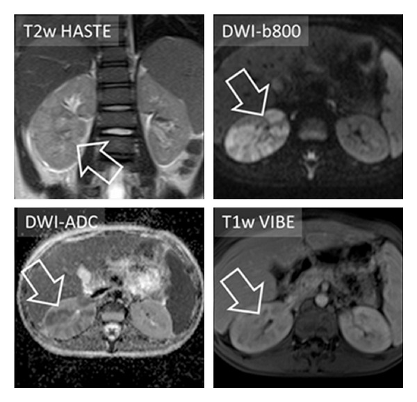
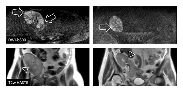
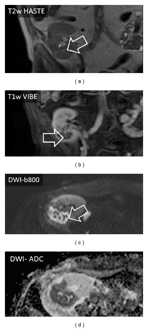
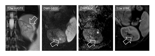
Twenty-one patients with suspicious increased signal intensity of the kidneys on DWI sequences and corresponding ADC decrease were identified. Sixty patients without clinical signs of renal infection served as a control group. All patients were examined with the following sequences: EPI-DWI (0/400/800 s/mm(2)), T2w HASTE, and T1w VIBE after intravenous injection of Gd-chelate. Confirmation of renal infection was established on the basis of clinical criteria. T1w and T2w images were assessed and compared to DWI for the presence of altered signal, and the degree of the visibility of pathology was graded on an ordinal three-point scale.
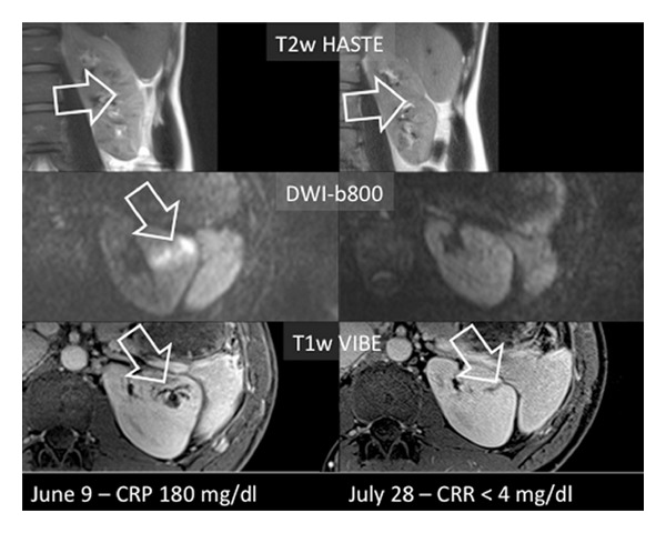
In all 21 patients with positive DWI findings a renal infection could be confirmed. T2w imaging and contrast-enhanced T1w imaging displayed obvious pathologic signal in 3/21 (14%) and 11/19 (58%) patients and slightly pathologic signal in 17/21 (81%) and 7/19 (37%), respectively. The median visibility score of 2 for the DWI and the T1w images was significantly higher than the score of 1 for the T2w imaging, P = 0.0001 (DWI versus T2w) and P = 0.078 (T1w versus T2w).
DWI of the kidneys seems to be highly sensitive for the detection of infections within the kidney.
评估扩散加权磁共振成像(DWI-MRI)在感染性肾病检测和评估中的应用。
确定21例在DWI序列上肾脏信号强度可疑增加且相应表观扩散系数(ADC)降低的患者。60例无肾脏感染临床症状的患者作为对照组。所有患者均采用以下序列进行检查:静脉注射钆螯合物后的EPI-DWI(0/400/800 s/mm²)、T2加权快速自旋回波(T2w HASTE)和T1加权容积内插屏气检查(T1w VIBE)。根据临床标准确诊肾脏感染。评估T1加权和T2加权图像,并与DWI图像对比观察信号改变情况,根据有序三分制对病变的可见程度进行分级。
所有21例DWI检查结果阳性的患者均确诊为肾脏感染。T2加权成像和对比增强T1加权成像分别在3/21(14%)和11/19(58%)的患者中显示明显病理信号,在17/21(81%)和7/19(37%)的患者中显示轻微病理信号。DWI和T1加权图像的中位可见度评分为2分,显著高于T2加权成像的1分,P = 0.0001(DWI与T2加权成像比较),P = 0.078(T1加权成像与T2加权成像比较)。
肾脏DWI对检测肾脏内感染似乎高度敏感。