• 文献检索
  • 文档翻译
  • 深度研究
  • 学术资讯
  • Suppr Zotero 插件Zotero 插件
  • 邀请有礼
  • 套餐&价格
  • 历史记录
应用&插件
Suppr Zotero 插件Zotero 插件浏览器插件Mac 客户端Windows 客户端微信小程序
定价
高级版会员购买积分包购买API积分包
服务
文献检索文档翻译深度研究API 文档MCP 服务
关于我们
关于 Suppr公司介绍联系我们用户协议隐私条款
关注我们

Suppr 超能文献

核心技术专利:CN118964589B侵权必究
粤ICP备2023148730 号-1Suppr @ 2026

文献检索

告别复杂PubMed语法,用中文像聊天一样搜索,搜遍4000万医学文献。AI智能推荐,让科研检索更轻松。

立即免费搜索

文件翻译

保留排版,准确专业,支持PDF/Word/PPT等文件格式,支持 12+语言互译。

免费翻译文档

深度研究

AI帮你快速写综述,25分钟生成高质量综述,智能提取关键信息,辅助科研写作。

立即免费体验

与对比增强计算机断层扫描相比,扩散加权磁共振成像在评估急性肾盂肾炎中的有效性。

Validity of diffusion-weighted magnetic resonance imaging in the evaluation of acute pyelonephritis in comparison with contrast-enhanced computed tomography.

作者信息

Sriman Rajasekaran, Venkatesh K, Mathew Cherian, Pankaj Mehta, Shankar Radhakrishnan

机构信息

Kovai Medical Center and Hospital (KMCH), Coimbatore, India.

Vinayaka Mission's Kirupananda Variyar Medical College (VMKVMCH), Salem, India.

出版信息

Pol J Radiol. 2020 Mar 14;85:e137-e143. doi: 10.5114/pjr.2020.93669. eCollection 2020.

DOI:10.5114/pjr.2020.93669
PMID:32322320
原文链接:https://pmc.ncbi.nlm.nih.gov/articles/PMC7172225/
Abstract

PURPOSE

Applications of diffusion-weighted magnetic resonance imaging outside the brain have gained increasing importance in recent years, and recent studies have shown the usage of diffusion-weighted (DW) imaging in diagnosing pyelonephritis based on renal cortical and medullary apparent diffusion coefficient (ADC) values. The aim of this study was to assess the validity of DW magnetic resonance (MR) imaging in comparison with contrast-enhanced computed tomography (CECT) in diagnosing pyelonephritis.

MATERIAL AND METHODS

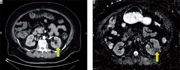
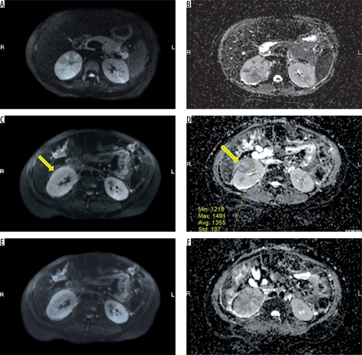
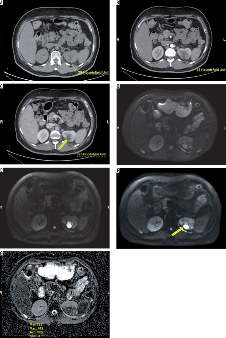
A cross-sectional observational study was conducted for a period of six months in a tertiary hospital in Coimbatore. All patients with clinical and laboratory diagnosis of acute pyelonephritis, who were referred for radiological imaging (CECT), were taken into the study. Out of 112 patients with a clinical and laboratorial diagnosis of acute pyelonephritis (APN), who underwent both DW MR and CECT, diagnosis of APN was made in 100 patients based on CECT, while in 12 cases the investigation (CECT) was negative. Finally, these 100 patients were included in the study. The validity of DW MR imaging in diagnosing APN was assessed by deriving sensitivity, specificity, and positive and negative predictive value in comparison with CECT findings.

RESULTS

The validity report of DW MR imaging in the detection of APN showed a very high sensitivity (96-100%) and specificity (86-90%) and very low false positives (6-10%) and negatives (< 5%), and it also showed that in the areas of affected renal parenchyma ADC values were consistently lower compared to unaffected renal parenchyma.

CONCLUSION

Based on the generated hypothesis, DW MR imaging of the kidneys seems to be highly sensitive and specific for the detection of focal or diffuse infections within the kidney in comparison with CECT.

摘要

目的

近年来,扩散加权磁共振成像在脑外的应用日益重要,近期研究表明扩散加权(DW)成像可基于肾皮质和髓质表观扩散系数(ADC)值用于诊断肾盂肾炎。本研究的目的是评估DW磁共振(MR)成像与对比增强计算机断层扫描(CECT)相比在诊断肾盂肾炎方面的有效性。

材料与方法

在哥印拜陀的一家三级医院进行了为期六个月的横断面观察性研究。所有临床和实验室诊断为急性肾盂肾炎且被转诊进行放射学成像(CECT)的患者均纳入研究。在112例临床和实验室诊断为急性肾盂肾炎(APN)且接受了DW MR和CECT检查的患者中,基于CECT诊断出100例APN,而12例检查(CECT)结果为阴性。最后,这100例患者被纳入研究。通过与CECT结果比较得出敏感性、特异性以及阳性和阴性预测值,来评估DW MR成像在诊断APN方面的有效性。

结果

DW MR成像检测APN的有效性报告显示出非常高的敏感性(96 - 100%)和特异性(86 - 90%),以及非常低的假阳性(6 - 10%)和假阴性(< 5%),并且还表明在受影响的肾实质区域,ADC值与未受影响的肾实质相比始终较低。

结论

基于所提出的假设,与CECT相比,肾脏的DW MR成像在检测肾脏内的局灶性或弥漫性感染方面似乎具有高度的敏感性和特异性。

https://cdn.ncbi.nlm.nih.gov/pmc/blobs/1885/7172225/24056df60652/PJR-85-40096-g003.jpg
https://cdn.ncbi.nlm.nih.gov/pmc/blobs/1885/7172225/4f5cb52b0e80/PJR-85-40096-g001.jpg
https://cdn.ncbi.nlm.nih.gov/pmc/blobs/1885/7172225/627c1a8a7065/PJR-85-40096-g002.jpg
https://cdn.ncbi.nlm.nih.gov/pmc/blobs/1885/7172225/24056df60652/PJR-85-40096-g003.jpg
https://cdn.ncbi.nlm.nih.gov/pmc/blobs/1885/7172225/4f5cb52b0e80/PJR-85-40096-g001.jpg
https://cdn.ncbi.nlm.nih.gov/pmc/blobs/1885/7172225/627c1a8a7065/PJR-85-40096-g002.jpg
https://cdn.ncbi.nlm.nih.gov/pmc/blobs/1885/7172225/24056df60652/PJR-85-40096-g003.jpg

相似文献

1
Validity of diffusion-weighted magnetic resonance imaging in the evaluation of acute pyelonephritis in comparison with contrast-enhanced computed tomography.与对比增强计算机断层扫描相比,扩散加权磁共振成像在评估急性肾盂肾炎中的有效性。
Pol J Radiol. 2020 Mar 14;85:e137-e143. doi: 10.5114/pjr.2020.93669. eCollection 2020.
2
Diffusion-weighted magnetic resonance imaging: new perspectives in the diagnostic pathway of non-complicated acute pyelonephritis.扩散加权磁共振成像:非复杂性急性肾盂肾炎诊断途径的新视角
Eur Radiol. 2013 Nov;23(11):3077-86. doi: 10.1007/s00330-013-2906-y. Epub 2013 Jun 8.
3
Acute pyelonephritis in transplanted kidneys: can diffusion-weighted magnetic resonance imaging be useful for diagnosis and follow-up?移植肾急性肾盂肾炎:弥散加权磁共振成像对诊断和随访是否有用?
Abdom Radiol (NY). 2016 Mar;41(3):531-7. doi: 10.1007/s00261-015-0618-2.
4
Follow-up of acute pyelonephritis: what causes the diffusion-weighted magnetic resonance imaging recovery to lag clinical recovery?急性肾盂肾炎的随访:是什么导致磁共振弥散加权成像恢复滞后于临床恢复?
Abdom Radiol (NY). 2018 Mar;43(3):639-646. doi: 10.1007/s00261-017-1242-0.
5
Diffusion-weighted imaging and apparent diffusion coefficient values versus contrast-enhanced MR imaging in the identification and characterisation of acute pyelonephritis.扩散加权成像和表观扩散系数值与对比增强磁共振成像在急性肾盂肾炎的识别和特征化中的比较。
Eur Radiol. 2013 Dec;23(12):3501-8. doi: 10.1007/s00330-013-2951-6. Epub 2013 Jul 26.
6
Role of diffusion-weighted MRI in acute pyelonephritis: a prospective study.弥散加权磁共振成像在急性肾盂肾炎中的作用:一项前瞻性研究。
Acta Radiol. 2015 Feb;56(2):244-9. doi: 10.1177/0284185114520862. Epub 2014 Jan 17.
7
Diffusion-weighted magnetic resonance imaging is more sensitive than dimercaptosuccinic acid scintigraphy in detecting parenchymal lesions in children with acute pyelonephritis: A prospective study.弥散加权磁共振成像比二巯丁二酸闪烁显像术更敏感,能检测出儿童急性肾盂肾炎的实质病变:一项前瞻性研究。
J Pediatr Urol. 2018 Jun;14(3):269.e1-269.e7. doi: 10.1016/j.jpurol.2018.02.014. Epub 2018 Mar 13.
8
Diffusion-weighted MR imaging of native and transplanted kidneys.弥散加权磁共振成像在天然和移植肾脏中的应用。
Radiology. 2011 Apr;259(1):25-38. doi: 10.1148/radiol.10092419.
9
Sensitivity of diffusion-weighted magnetic resonance imaging in the diagnosis of acute lacunar infarcts.扩散加权磁共振成像在急性腔隙性脑梗死诊断中的敏感性
J Formos Med Assoc. 2001 Jun;100(6):370-6.
10
Role of Diffusion Weighted MRI (DW-MR) in Detection of Satellite Lesions Not Detected with Multiphase CT Scans in Hepatoblastoma and Its Implications for Management.弥散加权磁共振成像(DW-MRI)在检测肝母细胞瘤多期 CT 扫描未检出卫星病灶中的作用及其对治疗的影响。
Indian J Pediatr. 2022 Oct;89(10):968-974. doi: 10.1007/s12098-021-04016-9. Epub 2022 Jan 8.

引用本文的文献

1
Diffusion-weighted MRI in the identification of renal parenchymal involvement in children with a first episode of febrile urinary tract infection.扩散加权磁共振成像在首次发热性尿路感染患儿肾实质受累情况识别中的应用
Front Radiol. 2024 Nov 21;4:1452902. doi: 10.3389/fradi.2024.1452902. eCollection 2024.
2
Contrast-Enhanced Ultrasound Follow-Up for Acute Pyelonephritis Patients.急性肾盂肾炎患者的超声造影随访
Healthcare (Basel). 2023 Nov 3;11(21):2899. doi: 10.3390/healthcare11212899.
3
Diffusion-Weighted MRI in the Genitourinary System.

本文引用的文献

1
Value of diffusion-weighted MR imaging for the detection of nephritis.磁共振扩散加权成像在肾炎检测中的价值。
ScientificWorldJournal. 2013 Nov 11;2013:348105. doi: 10.1155/2013/348105. eCollection 2013.
2
MRI and suspected acute pyelonephritis in children: comparison of diffusion-weighted imaging with gadolinium-enhanced T1-weighted imaging.MRI 与儿童疑似急性肾盂肾炎:扩散加权成像与钆增强 T1 加权成像的比较。
Eur Radiol. 2014 Jan;24(1):19-25. doi: 10.1007/s00330-013-2971-2. Epub 2013 Jul 25.
3
Noninvasive assessment of acute ureteral obstruction with diffusion-weighted MR imaging: a prospective study.
泌尿生殖系统的扩散加权磁共振成像
J Clin Med. 2022 Mar 30;11(7):1921. doi: 10.3390/jcm11071921.
4
Imaging of acute pelvic pain.急性盆腔痛的影像学表现。
Br J Radiol. 2021 Nov 1;94(1127):20210281. doi: 10.1259/bjr.20210281. Epub 2021 Sep 7.
5
CT and MRI in Urinary Tract Infections: A Spectrum of Different Imaging Findings.泌尿系统感染中的CT和MRI:一系列不同的影像学表现
Medicina (Kaunas). 2021 Jan 1;57(1):32. doi: 10.3390/medicina57010032.
磁共振扩散加权成像对急性输尿管梗阻的无创评估:一项前瞻性研究。
Radiology. 2009 Sep;252(3):721-8. doi: 10.1148/radiol.2523082090.
4
Diagnostic utility of diffusion-weighted MR imaging and apparent diffusion coefficient value for the diagnosis of adrenal tumors.磁共振扩散加权成像及表观扩散系数值在肾上腺肿瘤诊断中的应用价值
J Magn Reson Imaging. 2009 Jan;29(1):112-7. doi: 10.1002/jmri.21616.
5
Renal infarction in a patient with spontaneous dissection of segmental arteries: diffusion-weighted magnetic resonance imaging.节段性动脉自发性夹层患者的肾梗死:扩散加权磁共振成像
Am J Kidney Dis. 2008 Oct;52(4):788-91. doi: 10.1053/j.ajkd.2008.07.002. Epub 2008 Aug 26.
6
Diagnostic performance of low-dose CT for the detection of urolithiasis: a meta-analysis.低剂量CT检测尿路结石的诊断性能:一项荟萃分析
AJR Am J Roentgenol. 2008 Aug;191(2):396-401. doi: 10.2214/AJR.07.3414.
7
Comparison of 3.0-and 1.5-tesla diffusion-weighted imaging in the visibility of breast cancer.3.0特斯拉与1.5特斯拉扩散加权成像在乳腺癌可见性方面的比较。
Radiat Med. 2008 Jan;26(1):15-20. doi: 10.1007/s11604-007-0187-6. Epub 2008 Jan 31.
8
Functional evaluation of transplanted kidneys with diffusion-weighted and BOLD MR imaging: initial experience.磁共振扩散加权成像及血氧水平依赖性功能磁共振成像对移植肾的功能评估:初步经验
Radiology. 2006 Dec;241(3):812-21. doi: 10.1148/radiol.2413060103.
9
ADC measurement of abdominal organs and lesions using parallel imaging technique.使用并行成像技术对腹部器官和病变进行ADC测量。
AJR Am J Roentgenol. 2006 Dec;187(6):1521-30. doi: 10.2214/AJR.05.0778.
10
Normal and transplanted rat kidneys: diffusion MR imaging at 7 T.正常大鼠肾脏与移植大鼠肾脏:7T磁共振扩散成像
Radiology. 2004 Jun;231(3):702-9. doi: 10.1148/radiol.2313021587.