Stem Cell Res Ther. 2013;4(6):149. doi: 10.1186/scrt360.
Retrograde coronary venous infusion is a promising delivery method for cellular cardiomyoplasty. Poor cell retention is the major obstacle to the establishment of this method as the preferred route for cell delivery. Here, we explored whether magnetic targeting could enhance retrograde cell retention in a rat model of myocardial infarction.
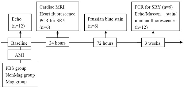
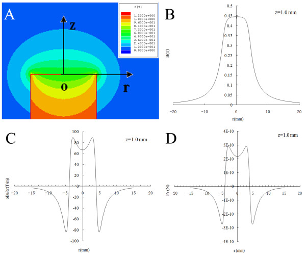
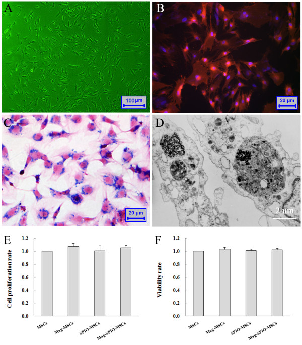
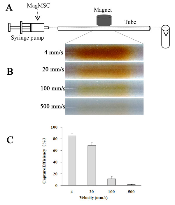
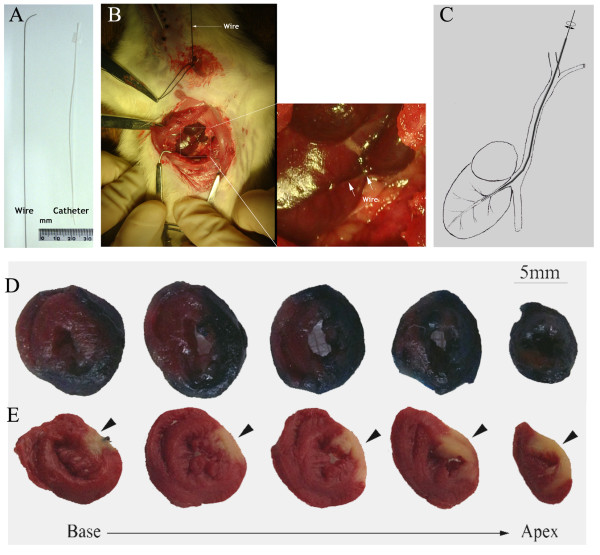
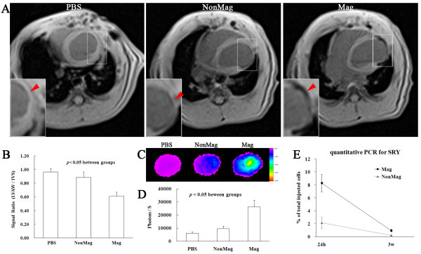
Rat mesenchymal stem cells were labeled with superparamagnetic oxide nanoparticles. The magnetic responsiveness of MSCs was observed while cells flowed through a tube that served as a model of blood vessels in a 0.6-Tesla magnetic field. In a Sprague–Dawley rat model of acute myocardial infarction, 1 × 10⁶ magnetic mesenchymal stem cells were transjugularly injected into the left cardiac vein while a 0.6-Tesla magnet was placed above the heart. The cardiac retention of transplanted cells was assessed by using quantitative Y chromosome-specific polymerase chain reaction, cardiac magnetic resonance imaging, and optical imaging. Cardiac function was measured by using echocardiography, and histologic analyses of infarct morphology and angiogenesis were obtained.
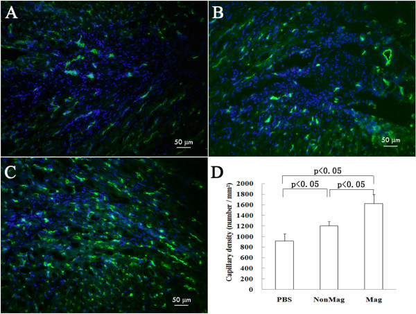
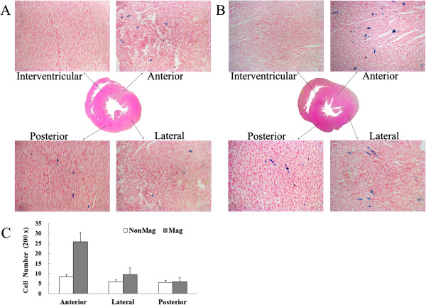
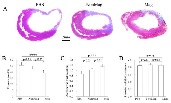
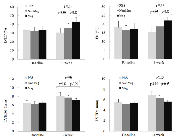
The flowing iron oxide-labeled mesenchymal stem cells were effectively attracted to the area where the magnet was positioned. Twenty-four hours after cellular retrocoronary delivery, magnetic targeting significantly increased the cardiac retention of transplanted cells by 2.73- to 2.87-fold. Histologic analyses showed that more transplanted cells were distributed in the anterior wall of the left ventricle. The enhanced cell engraftment persisted for at least 3 weeks, at which time, left ventricular remodeling was attenuated, and cardiac function benefit was improved.
These results suggest that magnetic targeting offers new perspectives for retrograde coronary venous delivery to enhance cell retention and subsequent functional benefit in heart diseases
逆行冠状静脉输注是细胞心肌成形术的一种很有前途的给药方法。细胞保留率低是该方法作为细胞输送首选途径的主要障碍。在这里,我们探讨了磁靶向是否可以增强心肌梗死大鼠模型中的逆行细胞保留。
用超顺磁氧化铁纳米颗粒标记大鼠间充质干细胞。当细胞在 0.6T 磁场中作为血管模型的管内流动时,观察 MSC 的磁响应。在急性心肌梗死的 Sprague-Dawley 大鼠模型中,将 1×10⁶ 个磁性间充质干细胞经颈内静脉逆行注射到左心静脉内,同时将 0.6T 磁铁置于心脏上方。通过定量 Y 染色体特异性聚合酶链反应、心脏磁共振成像和光学成像评估移植细胞的心脏保留情况。通过超声心动图测量心功能,并获得梗塞形态和血管生成的组织学分析。
流动的氧化铁标记间充质干细胞被有效地吸引到磁铁所在的区域。细胞逆行冠状动脉给药后 24 小时,磁靶向显著增加了移植细胞的心脏保留率,增加了 2.73-2.87 倍。组织学分析显示,更多的移植细胞分布在左心室前壁。增强的细胞植入持续至少 3 周,此时左心室重构得到减轻,心功能获益得到改善。
这些结果表明,磁靶向为逆行冠状静脉给药提供了新的视角,以增强细胞保留率,并在心脏病中获得随后的功能益处。