Department of Psychiatry, University of Alberta, Edmonton, AB, Canada.
J Neuroinflammation. 2013 Dec 13;10:152. doi: 10.1186/1742-2094-10-152.
Rasmussen's encephalitis (RE) is an inflammatory encephalopathy of unknown cause defined by seizures with progressive neurological disabilities. Herein, the pathogenesis of RE was investigated focusing on inflammasome activation in the brain.
Patients with RE at the University of Alberta, Edmonton, AB, Canada, were identified and analyzed by neuroimaging, neuropsychological, molecular, and pathological tools. Primary human microglia, astrocytes, and neurons were examined using RT-PCR, enzyme-linked immunosorbent assay (ELISA), and western blotting.
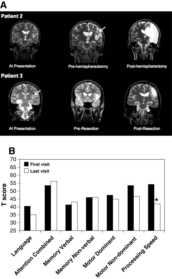
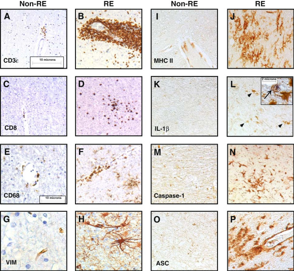
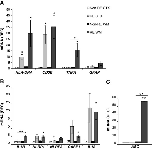
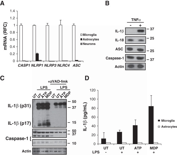
Four patients with RE were identified at the University of Alberta. Magnetic resonance imaging (MRI) disclosed increased signal intensities in cerebral white matter adjacent to cortical lesions of RE patients, accompanied by a decline in neurocognitive processing speed (P <0.05). CD3ϵ, HLA-DRA, and TNFα together with several inflammasome-associated genes (IL-1β, IL-18, NLRP1, NLRP3, and CASP1) showed increased transcript levels in RE brains compared to non-RE controls (n = 6; P <0.05). Cultured human microglia displayed expression of inflammasome-associated genes and responded to inflammasome activators by releasing IL-1β, which was inhibited by the caspase inhibitor, zVAD-fmk. Major histocompatibility complex (MHC) class II, IL-1β, caspase-1, and alanine/serine/cysteine (ASC) immunoreactivity were increased in RE brain tissues, especially in white matter myeloid cells, in conjunction with mononuclear cell infiltration and gliosis. Neuroinflammation in RE brains was present in both white matter and adjacent cortex with associated induction of inflammasome components, which was correlated with neuroimaging and neuropsychological deficits.
Inflammasome activation likely contributes to the disease process underlying RE and offers a mechanistic target for future therapeutic interventions.
拉森氏脑炎(RE)是一种病因不明的炎症性脑病,其特征是癫痫发作和进行性神经功能障碍。在此,我们通过神经影像学、神经心理学、分子和病理学工具,研究了 RE 的发病机制,重点关注了大脑中炎症小体的激活。
通过神经影像学、神经心理学、分子和病理学工具,在加拿大埃德蒙顿阿尔伯塔大学确定和分析了患有 RE 的患者。使用 RT-PCR、酶联免疫吸附测定(ELISA)和 Western blot 检测原代人小胶质细胞、星形胶质细胞和神经元。
在阿尔伯塔大学确定了 4 名患有 RE 的患者。磁共振成像(MRI)显示,RE 患者大脑皮质病变相邻的脑白质信号强度增加,同时神经认知处理速度下降(P <0.05)。与非 RE 对照组(n = 6;P <0.05)相比,RE 大脑中的 CD3ε、HLA-DRA 和 TNFα 以及几个炎症小体相关基因(IL-1β、IL-18、NLRP1、NLRP3 和 CASP1)的转录水平升高。培养的人小胶质细胞表达炎症小体相关基因,并通过释放 IL-1β 对炎症小体激活剂作出反应,该反应被半胱天冬酶抑制剂 zVAD-fmk 抑制。RE 脑组织中 MHC Ⅱ类、IL-1β、caspase-1 和丙氨酸/丝氨酸/半胱氨酸(ASC)免疫反应性增加,尤其是在白质髓细胞中,伴有单核细胞浸润和神经胶质增生。RE 大脑中的神经炎症存在于白质和邻近皮质中,伴有炎症小体成分的诱导,与神经影像学和神经心理学缺陷相关。
炎症小体的激活可能有助于 RE 发病机制,并为未来的治疗干预提供了一个机制靶点。