Che Yu, Bing Chen, Akhtar Javed, Tingting Zhao, Kezhou Yu, Rong Wang
Department of Nephrology, Provincial Hospital Affiliated to Shandong University, Shandong 250021, P, R, China.
J Transl Med. 2013 Dec 13;11:308. doi: 10.1186/1479-5876-11-308.
Arterial medial calcification (AMC) is frequent prevalence in patients with end stage renal disease. Evidence about hyperphosphatemia induced anabolic crosstalk between osteoblast and osteoclast in AMC of uremia is rare. Lanthanum carbonate as an orally administered phosphate-binding agent to reduce phosphate load and ameliorate AMC, but direct evidence is missing.
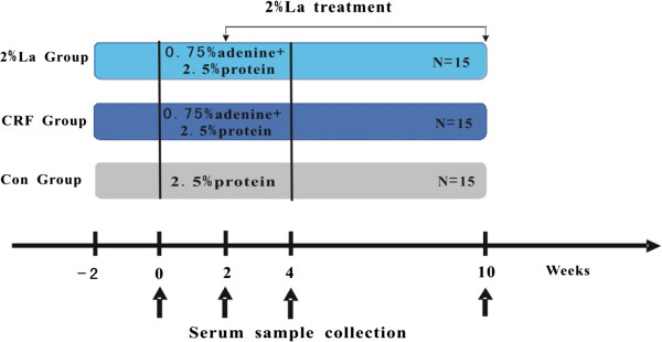
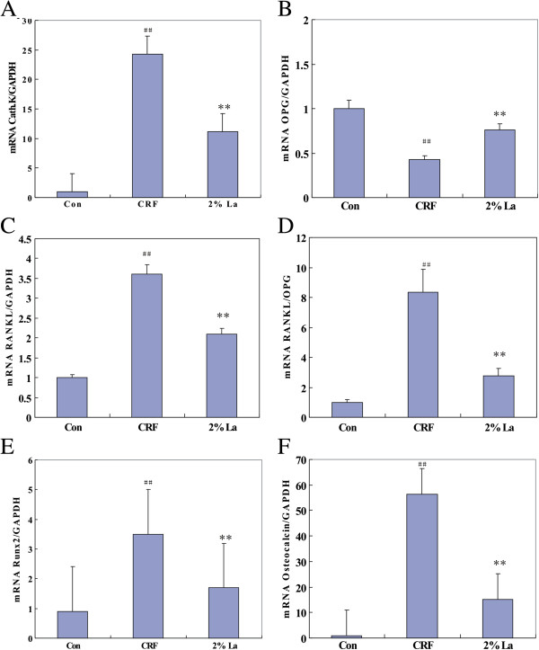
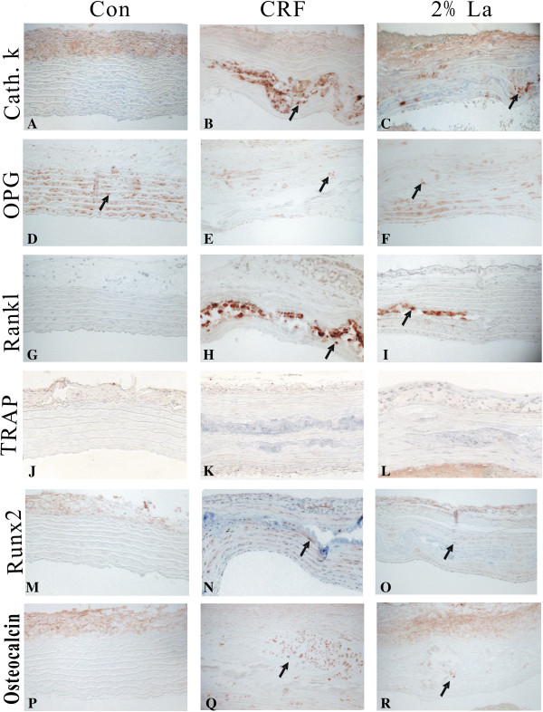
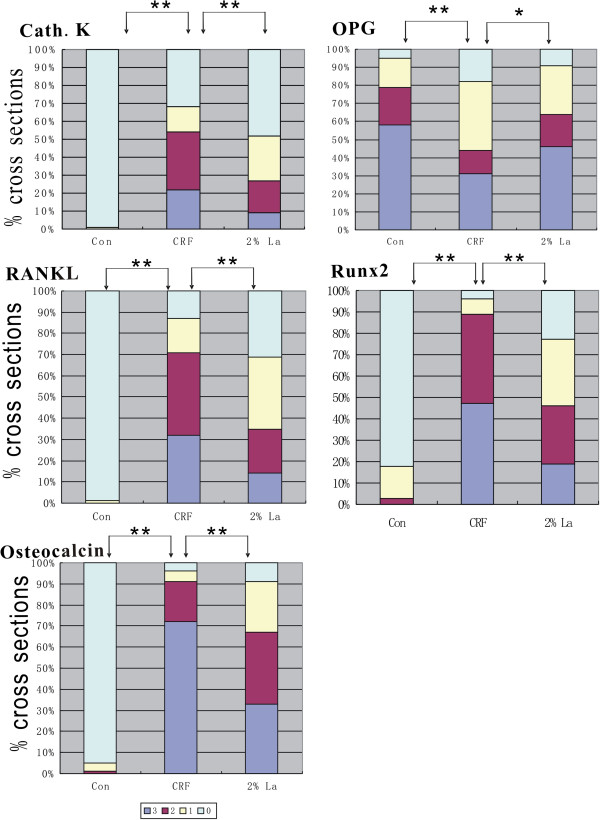
Detailed time-course studies were conducted of Sprague-Dawley rats fed with adenine and high phosphate diet to imitate the onset and progression of AMC of uremia. Calcification in great arteries was evaluated by VonKossa's and Masson's trichrome staining. Osteoblast (Runx2, Osteocalcin) and osteoclast (RANKL, Cathepsin K, TRAP) related genes were analyzed by Immunohistochemistry and qRT-PCR. Serum PTH, RANKL and OPG levels were detected by ELISA kit.
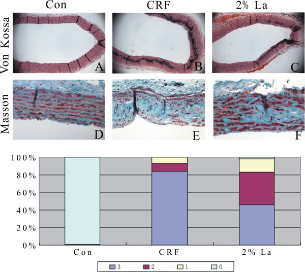
Serum phosphate was markedly increased in CRF group (6.94 ± 0.97 mmol/L) and 2%La group (5.12 ± 0.84 mmol/L) at week 4, while the latter group diminished significantly (2.92 ± 0.73 mmol/L vs CRF Group 3.48 ± 0.69, p < 0.01) at week 10. The rats that did not receive 2%La treatment had extensive von kossa staining for medial calcification in CRF group. In contrast, the rats in 2%La group just exhibit mild medial calcification. Inhibitory effect on progression of AMC was reflected by down regulated osteogenic genes and altered osteoclast-like genes. RANKL/OPG ratio in local calcification area was declined in 2%La group (vs CRF group, p <0.01), whereas marginal difference in serum among the three groups. In contrast to the robust expression of cathepsinK in calcified area, TRAP expression was not found.
Abnormal phosphate homeostasis, induction of osteogenic conversion and osteoclast suppression were contributed to the current mechanisms of uremia associated arterial medial calcification based on our studies. Beneficial effects of Lanthanum carbonate could be mainly due to the decreased phosphate retention and cross-talk between osteoblast and osteoclast-like cell, both of which can be the therapeutic target for uremia associated with AMC.
动脉中层钙化(AMC)在终末期肾病患者中普遍存在。关于高磷血症诱导尿毒症患者AMC中破骨细胞与成骨细胞之间合成代谢相互作用的证据很少。碳酸镧作为一种口服磷结合剂可减轻磷负荷并改善AMC,但缺乏直接证据。
对喂食腺嘌呤和高磷饮食的Sprague-Dawley大鼠进行详细的时间进程研究,以模拟尿毒症患者AMC的发生和发展。通过VonKossa染色和Masson三色染色评估大动脉钙化情况。采用免疫组织化学和qRT-PCR分析成骨细胞(Runx2、骨钙素)和破骨细胞(RANKL、组织蛋白酶K、抗酒石酸酸性磷酸酶)相关基因。用ELISA试剂盒检测血清甲状旁腺激素(PTH)、RANKL和骨保护素(OPG)水平。
第4周时,慢性肾衰竭(CRF)组(6.94±0.97 mmol/L)和2%碳酸镧组(5.12±0.84 mmol/L)血清磷显著升高,而第10周时,后一组血清磷显著降低(2.92±0.73 mmol/L vs CRF组3.48±0.69,p<0.01)。未接受2%碳酸镧治疗的大鼠,CRF组大动脉中层有广泛的VonKossa染色显示钙化。相比之下,2%碳酸镧组大鼠仅表现为轻度中层钙化。成骨基因下调和破骨样基因改变反映了碳酸镧对AMC进展的抑制作用。2%碳酸镧组局部钙化区域的RANKL/OPG比值下降(vs CRF组,p<0.01),而三组血清水平差异不显著。与组织蛋白酶K在钙化区域的强烈表达相反,未发现抗酒石酸酸性磷酸酶表达。
基于我们的研究,磷稳态异常、成骨转化诱导和破骨细胞抑制是尿毒症相关动脉中层钙化的现有机制。碳酸镧的有益作用可能主要归因于磷潴留减少以及成骨细胞与破骨样细胞之间的相互作用减少,这两者均可作为与AMC相关的尿毒症的治疗靶点。