Department of Biological Sciences, Vanderbilt University, Nashville, TN, USA.
Department of Ophthalmology, Emory University School of Medicine, Atlanta, GA, USA; Department of Pharmacology, Emory University School of Medicine, Atlanta, GA, USA.
Prog Retin Eye Res. 2014 Mar;39:58-76. doi: 10.1016/j.preteyeres.2013.12.001. Epub 2013 Dec 12.
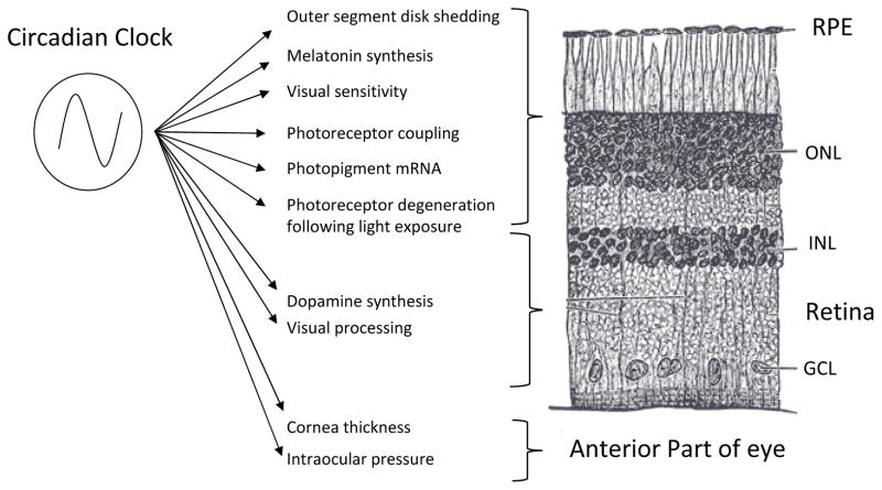
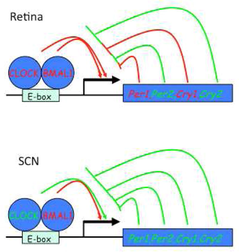
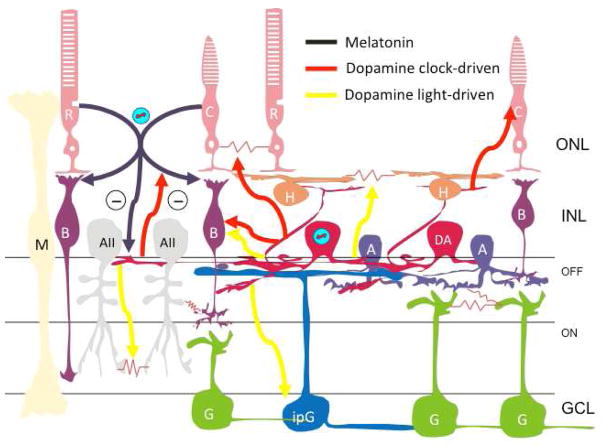
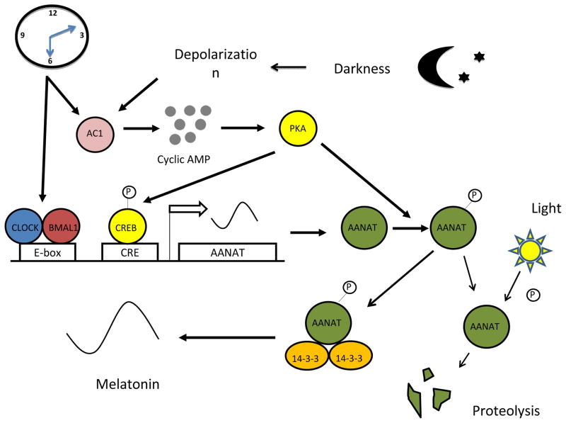
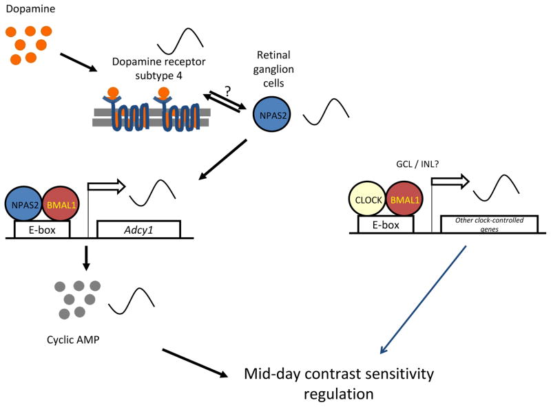
The retinal circadian system represents a unique structure. It contains a complete circadian system and thus the retina represents an ideal model to study fundamental questions of how neural circadian systems are organized and what signaling pathways are used to maintain synchrony of the different structures in the system. In addition, several studies have shown that multiple sites within the retina are capable of generating circadian oscillations. The strength of circadian clock gene expression and the emphasis of rhythmic expression are divergent across vertebrate retinas, with photoreceptors as the primary locus of rhythm generation in amphibians, while in mammals clock activity is most robust in the inner nuclear layer. Melatonin and dopamine serve as signaling molecules to entrain circadian rhythms in the retina and also in other ocular structures. Recent studies have also suggested GABA as an important component of the system that regulates retinal circadian rhythms. These transmitter-driven influences on clock molecules apparently reinforce the autonomous transcription-translation cycling of clock genes. The molecular organization of the retinal clock is similar to what has been reported for the SCN although inter-neural communication among retinal neurons that form the circadian network is apparently weaker than those present in the SCN, and it is more sensitive to genetic disruption than the central brain clock. The melatonin-dopamine system is the signaling pathway that allows the retinal circadian clock to reconfigure retinal circuits to enhance light-adapted cone-mediated visual function during the day and dark-adapted rod-mediated visual signaling at night. Additionally, the retinal circadian clock also controls circadian rhythms in disk shedding and phagocytosis, and possibly intraocular pressure. Emerging experimental data also indicate that circadian clock is also implicated in the pathogenesis of eye disease and compelling experimental data indicate that dysfunction of the retinal circadian system negatively impacts the retina and possibly the cornea and the lens.
视网膜昼夜节律系统代表了一种独特的结构。它包含一个完整的昼夜节律系统,因此视网膜是研究神经昼夜节律系统如何组织以及哪些信号通路用于维持系统中不同结构同步的理想模型。此外,多项研究表明,视网膜内的多个部位能够产生昼夜节律振荡。昼夜节律钟基因表达的强度和节律表达的重点在脊椎动物视网膜中是不同的,在两栖动物中,光感受器是节律产生的主要部位,而在哺乳动物中,生物钟活性在内核层最强。褪黑素和多巴胺作为信号分子,使视网膜和其他眼部结构的昼夜节律同步。最近的研究还表明 GABA 是调节视网膜昼夜节律的系统的重要组成部分。这些递质对生物钟分子的影响显然增强了生物钟基因的自主转录-翻译循环。视网膜时钟的分子组织与 SCN 中报道的相似,尽管形成昼夜节律网络的视网膜神经元之间的神经内通讯显然比 SCN 中的弱,而且比中枢大脑时钟更容易受到遗传破坏的影响。褪黑素-多巴胺系统是使视网膜昼夜节律时钟重新配置视网膜电路的信号通路,以增强白天适应光的锥体细胞介导的视觉功能,并在夜间适应暗的杆体细胞介导的视觉信号。此外,视网膜昼夜节律时钟还控制盘脱落和吞噬作用以及可能的眼内压的昼夜节律。新兴的实验数据还表明,昼夜节律时钟也与眼病的发病机制有关,有力的实验数据表明,视网膜昼夜节律系统的功能障碍会对视网膜产生负面影响,可能对角膜和晶状体也有影响。