Ruan Guo-Xiang, Allen Gregg C, Yamazaki Shin, McMahon Douglas G
Department of Biological Sciences, Vanderbilt University, Nashville, Tennessee, United States of America.
PLoS Biol. 2008 Oct 14;6(10):e249. doi: 10.1371/journal.pbio.0060249.
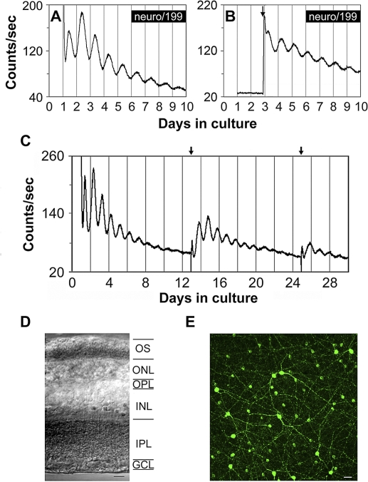
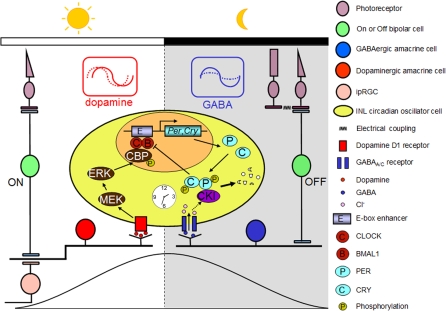
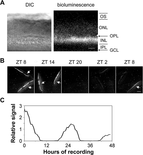
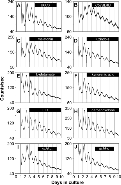
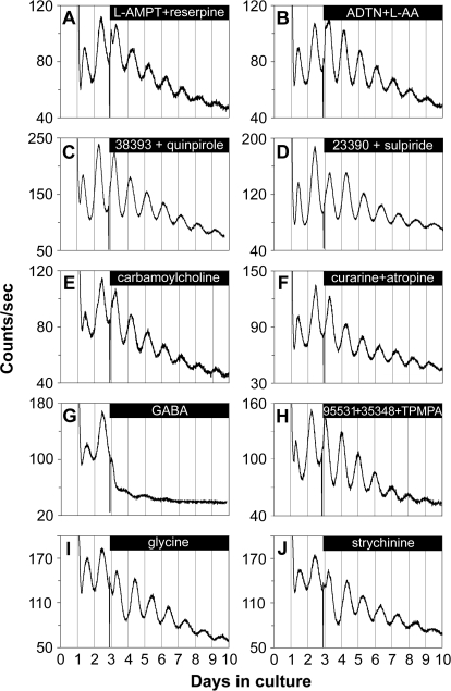
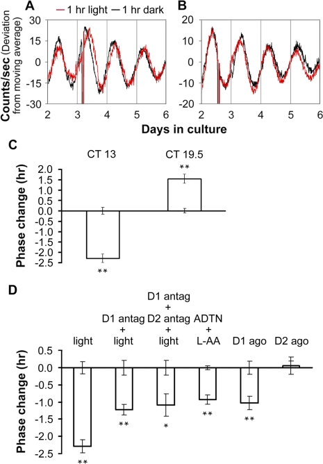
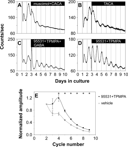
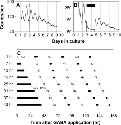
The influence of the mammalian retinal circadian clock on retinal physiology and function is widely recognized, yet the cellular elements and neural regulation of retinal circadian pacemaking remain unclear due to the challenge of long-term culture of adult mammalian retina and the lack of an ideal experimental measure of the retinal circadian clock. In the current study, we developed a protocol for long-term culture of intact mouse retinas, which allows retinal circadian rhythms to be monitored in real time as luminescence rhythms from a PERIOD2::LUCIFERASE (PER2::LUC) clock gene reporter. With this in vitro assay, we studied the characteristics and location within the retina of circadian PER2::LUC rhythms, the influence of major retinal neurotransmitters, and the resetting of the retinal circadian clock by light. Retinal PER2::LUC rhythms were routinely measured from whole-mount retinal explants for 10 d and for up to 30 d. Imaging of vertical retinal slices demonstrated that the rhythmic luminescence signals were concentrated in the inner nuclear layer. Interruption of cell communication via the major neurotransmitter systems of photoreceptors and ganglion cells (melatonin and glutamate) and the inner nuclear layer (dopamine, acetylcholine, GABA, glycine, and glutamate) did not disrupt generation of retinal circadian PER2::LUC rhythms, nor did interruption of intercellular communication through sodium-dependent action potentials or connexin 36 (cx36)-containing gap junctions, indicating that PER2::LUC rhythms generation in the inner nuclear layer is likely cell autonomous. However, dopamine, acting through D1 receptors, and GABA, acting through membrane hyperpolarization and casein kinase, set the phase and amplitude of retinal PER2::LUC rhythms, respectively. Light pulses reset the phase of the in vitro retinal oscillator and dopamine D1 receptor antagonists attenuated these phase shifts. Thus, dopamine and GABA act at the molecular level of PER proteins to play key roles in the organization of the retinal circadian clock.
哺乳动物视网膜生物钟对视网膜生理和功能的影响已得到广泛认可,但由于成年哺乳动物视网膜长期培养的挑战以及缺乏理想的视网膜生物钟实验测量方法,视网膜生物钟起搏的细胞成分和神经调节仍不清楚。在本研究中,我们开发了一种完整小鼠视网膜长期培养方案,该方案可通过PERIOD2::荧光素酶(PER2::LUC)生物钟基因报告基因的发光节律实时监测视网膜生物钟节律。通过这种体外测定方法,我们研究了昼夜节律性PER2::LUC节律在视网膜内的特征和位置、主要视网膜神经递质的影响以及光对视网膜生物钟的重置作用。从视网膜全层外植体中常规测量视网膜PER2::LUC节律10天,最长可达30天。垂直视网膜切片成像显示,节律性发光信号集中在内核层。通过光感受器和神经节细胞(褪黑素和谷氨酸)以及内核层(多巴胺、乙酰胆碱、GABA、甘氨酸和谷氨酸)的主要神经递质系统中断细胞通讯,以及通过钠依赖性动作电位或含连接蛋白36(cx36)的缝隙连接中断细胞间通讯,均未破坏视网膜昼夜节律性PER2::LUC节律的产生,这表明内核层中PER2::LUC节律的产生可能是细胞自主的。然而,通过D1受体起作用的多巴胺和通过膜超极化和酪蛋白激酶起作用的GABA分别设定了视网膜PER2::LUC节律的相位和幅度。光脉冲重置了体外视网膜振荡器的相位,多巴胺D1受体拮抗剂减弱了这些相位变化。因此,多巴胺和GABA在PER蛋白的分子水平上发挥作用,在视网膜生物钟的组织中起关键作用。