Babalola Oe, Adu A, Akano Ao
Bingham University, New Karu, Nassarawa State, Abuja, Nigeria.
Department of Ophthalmology, Rachel Eye Center, Garki Phase II, Abuja, Nigeria.
Clin Ophthalmol. 2013;7:2275-9. doi: 10.2147/OPTH.S52690. Epub 2013 Dec 3.
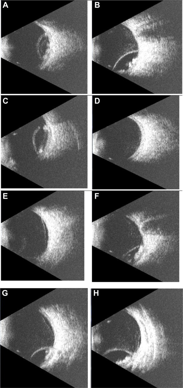
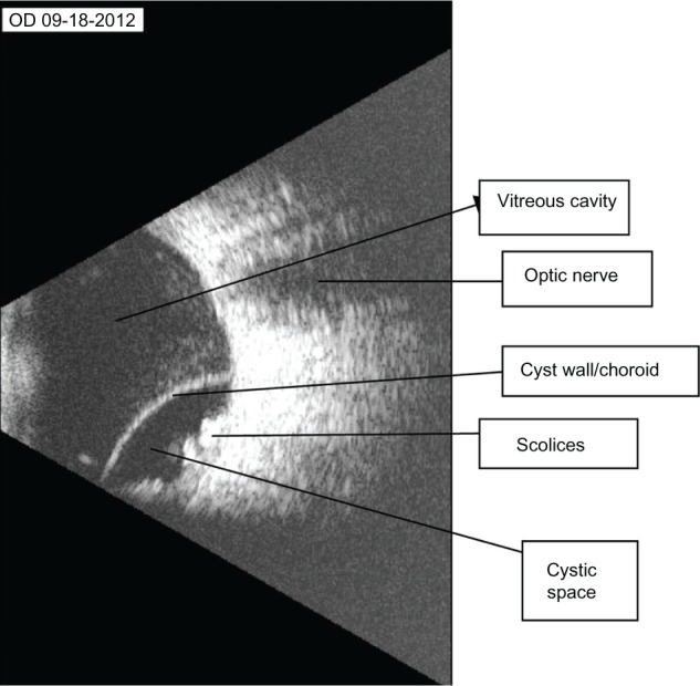
We report the case of a 32-year-old man suffering from intraocular cysticercosis, with special emphasis on the use of B-scan ultrasound in the diagnosis and management of the condition. An 8000 B-Scan Scanmate was used to obtain the ultrasound images. The patient had worked on a pig farm a few years before presentation. He presented with shadows seen in the right eye. Binocular indirect ophthalmoscopy revealed that he had a choroidal detachment in the right eye inferotemporally. B-scan ultrasound revealed a subretinal subchoroidal cyst with a thick wall containing well defined intracystic echogenic entities representing scolices, and an associated retinal detachment. These findings appear to be pathognomonic. Excision of the cyst through a trans-scleral approach revealed a yellowish serous fluid, with scolices of cysticercus later confirmed histologically. B-scan ultrasound is extremely useful in the diagnosis of ocular cysticercosis and the findings can be pathognomonic.
我们报告了一例32岁患有眼内囊尾蚴病的男性病例,特别强调了B超在该疾病诊断和治疗中的应用。使用8000型B超Scanmate获取超声图像。该患者在就诊前几年曾在养猪场工作。他右眼出现阴影。双目间接检眼镜检查发现其右眼颞下象限脉络膜脱离。B超显示视网膜下脉络膜下有一个囊肿,囊肿壁增厚,囊内有明确的呈头节状的强回声实体,伴有视网膜脱离。这些表现似乎具有特征性。通过经巩膜途径切除囊肿,可见淡黄色浆液性液体,囊尾蚴头节经组织学检查得以证实。B超在眼囊尾蚴病的诊断中极其有用,其表现具有特征性。