• 文献检索
  • 文档翻译
  • 深度研究
  • 学术资讯
  • Suppr Zotero 插件Zotero 插件
  • 邀请有礼
  • 套餐&价格
  • 历史记录
应用&插件
Suppr Zotero 插件Zotero 插件浏览器插件Mac 客户端Windows 客户端微信小程序
定价
高级版会员购买积分包购买API积分包
服务
文献检索文档翻译深度研究API 文档MCP 服务
关于我们
关于 Suppr公司介绍联系我们用户协议隐私条款
关注我们

Suppr 超能文献

核心技术专利:CN118964589B侵权必究
粤ICP备2023148730 号-1Suppr @ 2026

文献检索

告别复杂PubMed语法,用中文像聊天一样搜索,搜遍4000万医学文献。AI智能推荐,让科研检索更轻松。

立即免费搜索

文件翻译

保留排版,准确专业,支持PDF/Word/PPT等文件格式,支持 12+语言互译。

免费翻译文档

深度研究

AI帮你快速写综述,25分钟生成高质量综述,智能提取关键信息,辅助科研写作。

立即免费体验

用于电子显微镜的金属投影法。

Metal shadowing for electron microscopy.

作者信息

Hendricks Gregory M

机构信息

Core Electron Microscopy Facility, Department of Cell Biology, University of Massachusetts Medical School, Worcester, MA, USA.

出版信息

Methods Mol Biol. 2014;1117:73-93. doi: 10.1007/978-1-62703-776-1_5.

DOI:10.1007/978-1-62703-776-1_5
PMID:24357360
原文链接:https://pmc.ncbi.nlm.nih.gov/articles/PMC7120032/
Abstract

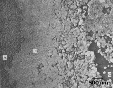
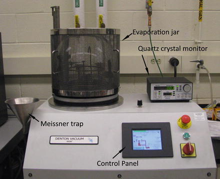
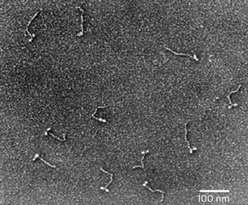
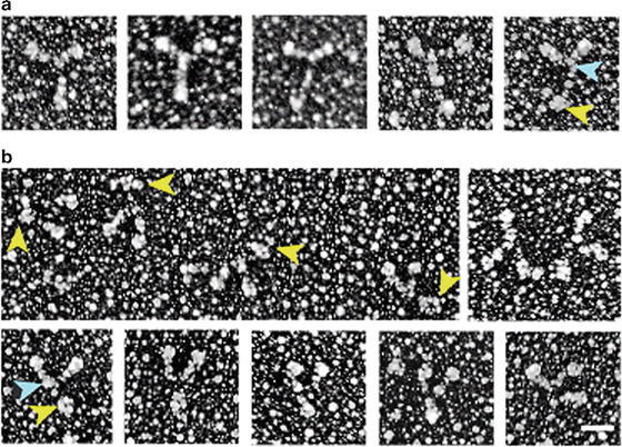
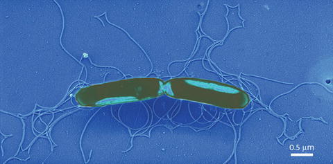
Metal shadowing of bacteria, viruses, isolated molecules, and macromolecular assemblies is another high-resolution method for observing the ultrastructure of biological specimens. The actual procedure for producing a metal shadow is relatively simple; a heavy metal is evaporated from a source at an oblique angle to the specimen. The metal atoms pile up on the surfaces that face the source, but the surfaces away from the source are shielded and receive little metal deposit, creating a "shadow." However, the process of producing biological specimens that are suitable for metal shadowing can be very complex. There are a whole host of specimen preparation techniques that can precede metal shadowing, and all provide superior preservation in comparison to air drying, a required step in negative staining procedures. The physical forces present during air drying (i.e., surface tension of the water-air interface) will literally crush most biological specimens as they dry. In this chapter I explain the development of and procedures for the production of biological specimens from macromolecular assemblies (e.g., DNA and RNA), purified isolated molecules (e.g., proteins), and isolated viruses and bacteria preparations suitable for metal shadowing. A variation on this basic technique is to rotate the specimen during the metal deposition to produce a high-resolution three-dimensional rendering of the specimen.

摘要

对细菌、病毒、分离出的分子以及大分子聚集体进行金属投影是观察生物标本超微结构的另一种高分辨率方法。产生金属投影的实际过程相对简单;重金属从一个源以倾斜角度蒸发到标本上。金属原子堆积在面对源的表面上,但远离源的表面被屏蔽,几乎没有金属沉积,从而形成一个“投影”。然而,制备适合金属投影的生物标本的过程可能非常复杂。在金属投影之前有一整套标本制备技术,并且与负染色程序中必需的空气干燥步骤相比,所有这些技术都能提供更好的保存效果。空气干燥过程中存在的物理力(即水 - 空气界面的表面张力)在标本干燥时实际上会压碎大多数生物标本。在本章中,我将解释从大分子聚集体(如DNA和RNA)、纯化的分离分子(如蛋白质)以及适合金属投影的分离病毒和细菌制剂制备生物标本的发展过程和程序。这种基本技术的一个变体是在金属沉积过程中旋转标本,以生成标本的高分辨率三维图像。

https://cdn.ncbi.nlm.nih.gov/pmc/blobs/b470/7120032/4861b6c03bbe/300616_3_En_5_Fig9_HTML.jpg
https://cdn.ncbi.nlm.nih.gov/pmc/blobs/b470/7120032/5e9071ebedda/300616_3_En_5_Fig1_HTML.jpg
https://cdn.ncbi.nlm.nih.gov/pmc/blobs/b470/7120032/7859bda6164d/300616_3_En_5_Fig2_HTML.jpg
https://cdn.ncbi.nlm.nih.gov/pmc/blobs/b470/7120032/6929a89326cc/300616_3_En_5_Fig3_HTML.jpg
https://cdn.ncbi.nlm.nih.gov/pmc/blobs/b470/7120032/c612954bbf44/300616_3_En_5_Fig4_HTML.jpg
https://cdn.ncbi.nlm.nih.gov/pmc/blobs/b470/7120032/064d6058df9a/300616_3_En_5_Fig5_HTML.jpg
https://cdn.ncbi.nlm.nih.gov/pmc/blobs/b470/7120032/351a60eee8c4/300616_3_En_5_Fig6_HTML.jpg
https://cdn.ncbi.nlm.nih.gov/pmc/blobs/b470/7120032/f67fad0f784c/300616_3_En_5_Fig7_HTML.jpg
https://cdn.ncbi.nlm.nih.gov/pmc/blobs/b470/7120032/36158bf3e106/300616_3_En_5_Fig8_HTML.jpg
https://cdn.ncbi.nlm.nih.gov/pmc/blobs/b470/7120032/4861b6c03bbe/300616_3_En_5_Fig9_HTML.jpg

相似文献

1
Metal shadowing for electron microscopy.用于电子显微镜的金属投影法。
Methods Mol Biol. 2014;1117:73-93. doi: 10.1007/978-1-62703-776-1_5.
2
Sputter shadowing.
J Ultrastruct Res. 1984 May;87(2):97-105. doi: 10.1016/s0022-5320(84)80069-6.
3
Image interpretation of metal shadowed specimens with the aid of Monte Carlo simulation of the shadowing process.借助阴影过程的蒙特卡罗模拟对金属阴影标本进行图像解释。
Scanning Microsc Suppl. 1988;2:379-86.
4
Preparation of viral samples within biocontainment for ultrastructural analysis: Utilization of an innovative processing capsule for negative staining.在生物安全防护设施内制备用于超微结构分析的病毒样本:使用创新型处理胶囊进行负染色
J Virol Methods. 2016 Dec;238:70-76. doi: 10.1016/j.jviromet.2016.10.005. Epub 2016 Oct 14.
5
Metal shadowing and decoration in electron microscopy of biological macromolecules.
Ann N Y Acad Sci. 1986;483:57-76. doi: 10.1111/j.1749-6632.1986.tb34497.x.
6
Bidirectional shadowing in freeze-etching.
J Microsc. 1983 Nov;132(Pt 2):171-8. doi: 10.1111/j.1365-2818.1983.tb04269.x.
7
Correlation of surface topography of metal-shadowed specimens with their negatively stained reconstructions.
Ultramicroscopy. 1985;16(3-4):436-50. doi: 10.1016/0304-3991(85)90110-x.
8
Improved handling of structural fragile cell-biological specimens during electron microscopic preparation by the exchange method.通过交换法在电子显微镜制备过程中对结构脆弱的细胞生物学标本进行改进处理。
J Microsc. 1980 Apr;118(4):429-41. doi: 10.1111/j.1365-2818.1980.tb00292.x.
9
Ultrahigh-resolution scanning electron microscopy and its application to medical researches.
Rhinol Suppl. 1992;14:8-13.
10
Determining the structure of biological macromolecules by transmission electron microscopy, single particle analysis and 3D reconstruction.通过透射电子显微镜、单颗粒分析和三维重建确定生物大分子的结构。
Prog Biophys Mol Biol. 2001;75(3):121-64. doi: 10.1016/s0079-6107(01)00004-9.

引用本文的文献

1
Fine structure of the vaccinia virion determined by controlled degradation and immunolocalization.通过可控降解和免疫定位确定的痘苗病毒粒子的精细结构
Virology. 2015 Jan 15;475:204-18. doi: 10.1016/j.virol.2014.11.020. Epub 2014 Dec 8.

本文引用的文献

1
Modern uses of electron microscopy for detection of viruses.电子显微镜在病毒检测中的现代应用。
Clin Microbiol Rev. 2009 Oct;22(4):552-63. doi: 10.1128/CMR.00027-09.
2
Probing the structure of the SARS coronavirus using scanning electron microscopy.利用扫描电子显微镜探究严重急性呼吸综合征冠状病毒的结构。
Antivir Ther. 2004 Apr;9(2):287-9.
3
Myosin V: regulation by calcium, calmodulin, and the tail domain.肌球蛋白V:受钙、钙调蛋白和尾部结构域调控。
J Cell Biol. 2004 Mar 15;164(6):877-86. doi: 10.1083/jcb.200310065. Epub 2004 Mar 8.
4
The egg white inhibitor of influenza virus hemagglutination. II. Electron microscopy of the inhibitor.
J Biol Chem. 1950 Aug;185(2):681-8.
5
Simultaneous evaporation of platinum and carbon for possible use in high-resolution shadow-casting for the electron microscope.铂和碳的同时蒸发,可能用于电子显微镜的高分辨率投影。
Nature. 1958 Mar 29;181(4613):875-7. doi: 10.1038/181875a0.
6
Genetic identification and characterization of a novel virus related to human hepatitis E virus from chickens with hepatitis-splenomegaly syndrome in the United States.从美国患有肝脾肿大综合征的鸡中分离出一种与人类戊型肝炎病毒相关的新型病毒的基因鉴定与特征分析。
J Gen Virol. 2001 Oct;82(Pt 10):2449-2462. doi: 10.1099/0022-1317-82-10-2449.
7
Quick and easy spreading technique for electron microscopy of DNA.
Biotechniques. 1995 Jun;18(6):946-7.
8
Ultrastructure of the cell envelope layers and surface details of Legionella pneumophila.
J Gen Microbiol. 1982 Jul;128(7):1547-57. doi: 10.1099/00221287-128-7-1547.
9
The inside and outside of gap-junction membranes visualized by deep etching.通过深度蚀刻观察到的间隙连接膜的内部和外部。
Cell. 1982 Sep;30(2):395-406. doi: 10.1016/0092-8674(82)90237-9.
10
Preparation of single molecules and supramolecular complexes for high-resolution metal shadowing.用于高分辨率金属投影的单分子和超分子复合物的制备。
J Ultrastruct Res. 1983 Jun;83(3):319-34. doi: 10.1016/s0022-5320(83)90139-9.