Foroutan Parastou, Kreahling Jenny M, Morse David L, Grove Olya, Lloyd Mark C, Reed Damon, Raghavan Meera, Altiok Soner, Martinez Gary V, Gillies Robert J
Department of Cancer Imaging and Metabolism, H. Lee Moffitt Cancer Center & Research Institute, Tampa, Florida, United States of America.
Experimental Therapeutics Program, H. Lee Moffitt Cancer Center & Research Institute, Tampa, Florida, United States of America.
PLoS One. 2013 Dec 16;8(12):e82875. doi: 10.1371/journal.pone.0082875. eCollection 2013.
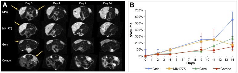
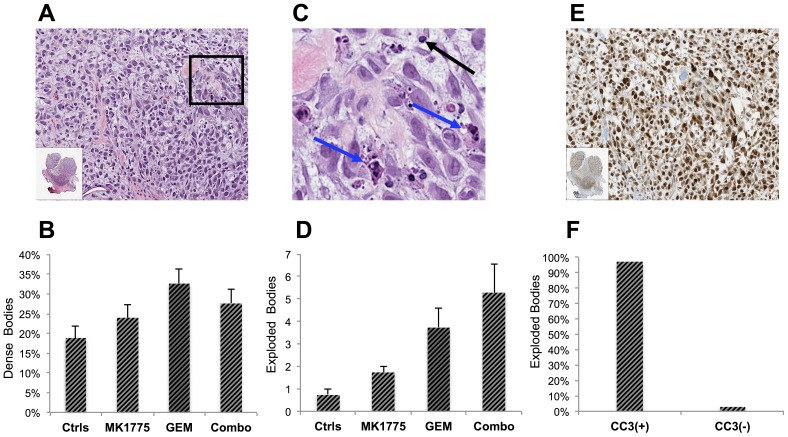
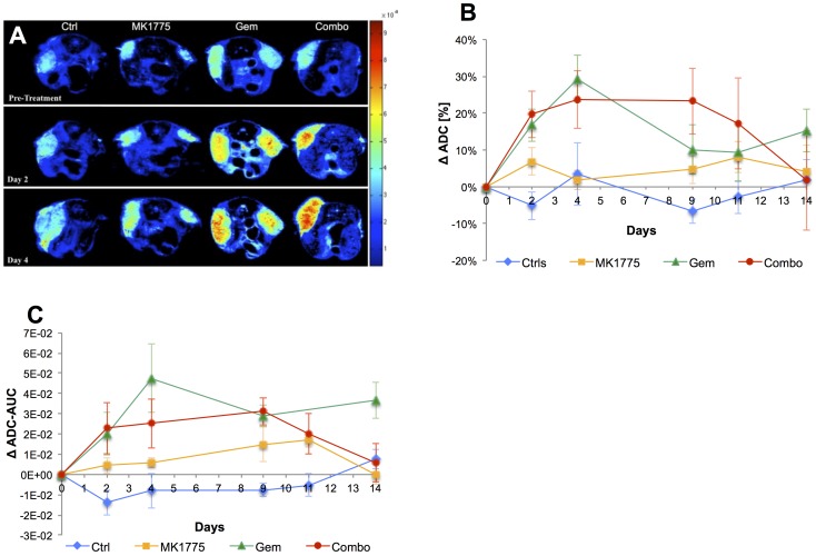
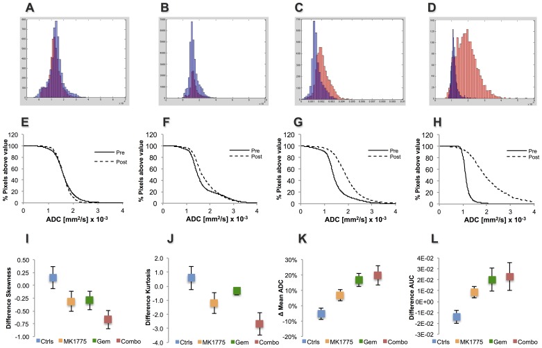
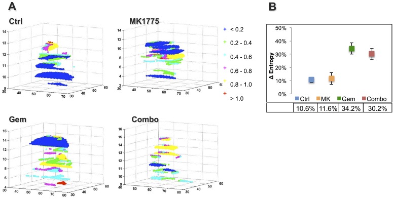
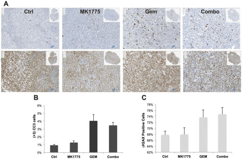
Combinations of targeted drugs have been employed to treat sarcomas, however, response rates have not improved notably, therefore emphasizing the need for novel treatments. In addition, imaging approaches to assess therapeutic response is lacking, as currently measurable indices, such as volume and/or diameter, do not accurately correlate with changes in tumor biology. In this study, quantitative and profound analyses of magnetic resonance imaging (MRI) were developed to evaluate these as imaging biomarkers for MK1775 and Gem in an osteosarcoma xenotransplant model at early time-points following treatment. Notably, we showed that Gem and Gem+MK1775 groups had significantly inhibited tumor growth by day 4, which was presaged by elevations in mean ADC by 24 hours post treatment. Significant differences were also observed at later time points for the Gem+MK1775 combination and MK1775 therapy. ADC distribution and entropy (randomness of ADC values) were also elevated by 24 hours following therapy. Immunohistochemistry demonstrated that these treatment-related increases in ADC correlated with apoptosis and observed cell condensations (dense- and exploded bodies). These findings underline the role of ADC as a quantitative imaging biomarker for therapy-induced response and show promising clinical relevance in the sarcoma patient population.
靶向药物联合已被用于治疗肉瘤,然而,缓解率并未显著提高,因此凸显了新型治疗方法的必要性。此外,目前缺乏评估治疗反应的影像学方法,因为目前可测量的指标,如体积和/或直径,与肿瘤生物学变化并无准确关联。在本研究中,我们开展了磁共振成像(MRI)的定量和深入分析,以评估其作为骨肉瘤异种移植模型中MK1775和吉西他滨(Gem)在治疗后早期时间点的成像生物标志物。值得注意的是,我们发现,到第4天时,吉西他滨组和吉西他滨+MK1775组的肿瘤生长受到显著抑制,这在治疗后24小时平均表观扩散系数(ADC)升高时就已有所预示。在后续时间点,吉西他滨+MK1775联合治疗组和MK1775治疗组也观察到了显著差异。治疗后24小时,ADC分布和熵(ADC值的随机性)也有所升高。免疫组织化学显示,这些与治疗相关的ADC升高与细胞凋亡以及观察到的细胞凝聚(致密体和爆炸体)相关。这些发现强调了ADC作为治疗诱导反应的定量成像生物标志物的作用,并在肉瘤患者群体中显示出有前景的临床相关性。