• 文献检索
  • 文档翻译
  • 深度研究
  • 学术资讯
  • Suppr Zotero 插件Zotero 插件
  • 邀请有礼
  • 套餐&价格
  • 历史记录
应用&插件
Suppr Zotero 插件Zotero 插件浏览器插件Mac 客户端Windows 客户端微信小程序
定价
高级版会员购买积分包购买API积分包
服务
文献检索文档翻译深度研究API 文档MCP 服务
关于我们
关于 Suppr公司介绍联系我们用户协议隐私条款
关注我们

Suppr 超能文献

核心技术专利:CN118964589B侵权必究
粤ICP备2023148730 号-1Suppr @ 2026

文献检索

告别复杂PubMed语法,用中文像聊天一样搜索,搜遍4000万医学文献。AI智能推荐,让科研检索更轻松。

立即免费搜索

文件翻译

保留排版,准确专业,支持PDF/Word/PPT等文件格式,支持 12+语言互译。

免费翻译文档

深度研究

AI帮你快速写综述,25分钟生成高质量综述,智能提取关键信息,辅助科研写作。

立即免费体验

MRI 上手动和半自动骨肉瘤分割具有高度相似性。

Manual and semiautomatic segmentation of bone sarcomas on MRI have high similarity.

机构信息

Departamento de Imagens Médicas, Hematologia e Oncologia Clínica, Faculdade de Medicina de Ribeirão Preto, Universidade de São Paulo, Ribeirão Preto, SP, Brasil.

Laboratório de Pesquisa em Imagens Musculoesqueléticas, Faculdade de Medicina de Ribeirão Preto, Universidade de São Paulo, Ribeirão Preto, SP, Brasil.

出版信息

Braz J Med Biol Res. 2020 Jan 31;53(2):e8962. doi: 10.1590/1414-431X20198962. eCollection 2020.

DOI:10.1590/1414-431X20198962
PMID:32022102
原文链接:https://pmc.ncbi.nlm.nih.gov/articles/PMC6993358/
Abstract

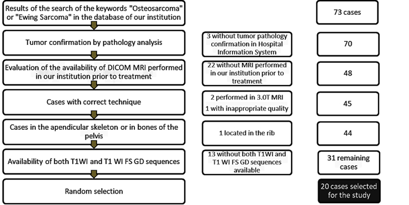
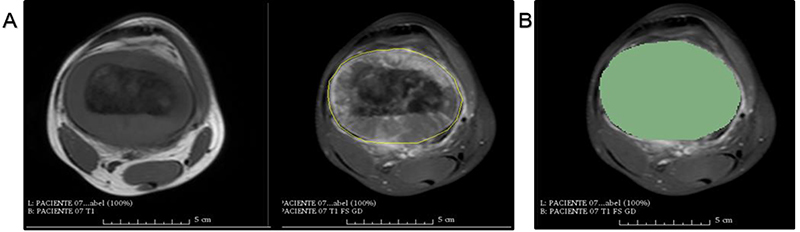
The aims of this study were to evaluate the intra- and interobserver reproducibility of manual segmentation of bone sarcomas in magnetic resonance imaging (MRI) studies and to compare manual and semiautomatic segmentation methods. This retrospective study included twelve osteosarcoma and eight Ewing sarcoma MRI studies performed prior to any therapeutic intervention. All cases were histopathologically confirmed. Three radiologists used 3D-Slicer software to perform manual segmentation of bone sarcomas in a blinded and independent manner. One radiologist segmented manually and also performed semiautomatic segmentation with the GrowCut tool. Segmentation exercises were timed for comparison. The dice similarity coefficient (DSC) and Hausdorff distance (HD) were used to evaluate similarity between the segmentation results and further statistical analyses were performed to compare DSC, HD, and volumetric results. Manual segmentation was reproducible with intraobserver DSC varying from 0.83 to 0.97 and HD from 3.37 to 28.73 mm. Interobserver DSC of manual segmentation showed variation from 0.73 to 0.97 and HD from 3.93 to 33.40 mm. Semiautomatic segmentation compared to manual segmentation resulted in DSCs of 0.71-0.96 and HDs of 5.38-31.54 mm. Semiautomatic segmentation required significantly less time compared to manual segmentation (P value ≤0.05). Among all situations compared, tumor volumetry did not show significant statistical differences (P value >0.05). We found excellent intra- and interobserver agreement for manual segmentation of osteosarcoma and Ewing sarcoma. There was high similarity between manual and semiautomatic segmentation, with a significant reduction of segmentation time using the semiautomatic method.

摘要

本研究旨在评估磁共振成像(MRI)研究中骨肉瘤手动分割的观察者内和观察者间可重复性,并比较手动和半自动分割方法。这项回顾性研究包括 12 例骨肉瘤和 8 例尤文肉瘤的 MRI 研究,这些研究均在任何治疗干预之前进行。所有病例均经组织病理学证实。三位放射科医生使用 3D-Slicer 软件以盲法和独立的方式进行骨肉瘤的手动分割。一位放射科医生手动分割,并使用 GrowCut 工具进行半自动分割。分割练习的用时用于比较。使用骰子相似系数(DSC)和 Hausdorff 距离(HD)评估分割结果之间的相似性,并进一步进行统计学分析以比较 DSC、HD 和体积结果。手动分割具有可重复性,观察者内 DSC 从 0.83 到 0.97,HD 从 3.37 到 28.73mm。手动分割的观察者间 DSC 变化范围为 0.73 到 0.97,HD 为 3.93 到 33.40mm。半自动分割与手动分割相比,DSC 为 0.71-0.96,HD 为 5.38-31.54mm。半自动分割比手动分割明显需要更少的时间(P 值≤0.05)。在所有比较的情况下,肿瘤体积均无显著统计学差异(P 值>0.05)。我们发现骨肉瘤和尤文肉瘤的手动分割具有极好的观察者内和观察者间一致性。手动和半自动分割之间具有高度相似性,半自动方法显著减少了分割时间。

https://cdn.ncbi.nlm.nih.gov/pmc/blobs/659c/6993358/1a943f56d908/1414-431X-bjmbr-53-2-e8962-gf003.jpg
https://cdn.ncbi.nlm.nih.gov/pmc/blobs/659c/6993358/7964a91ac0b5/1414-431X-bjmbr-53-2-e8962-gf001.jpg
https://cdn.ncbi.nlm.nih.gov/pmc/blobs/659c/6993358/77d4e17a215e/1414-431X-bjmbr-53-2-e8962-gf002.jpg
https://cdn.ncbi.nlm.nih.gov/pmc/blobs/659c/6993358/1a943f56d908/1414-431X-bjmbr-53-2-e8962-gf003.jpg

相似文献

1
Manual and semiautomatic segmentation of bone sarcomas on MRI have high similarity.MRI 上手动和半自动骨肉瘤分割具有高度相似性。
Braz J Med Biol Res. 2020 Jan 31;53(2):e8962. doi: 10.1590/1414-431X20198962. eCollection 2020.
2
Manual versus semiautomatic segmentation of soft-tissue sarcomas on magnetic resonance imaging: evaluation of similarity and comparison of segmentation times.磁共振成像中软组织肉瘤的手动分割与半自动分割:相似性评估及分割时间比较
Radiol Bras. 2021 May-Jun;54(3):155-164. doi: 10.1590/0100-3984.2020.0028.
3
Semiautomatic quantification of carotid plaque volume with three-dimensional ultrasound imaging.利用三维超声成像对颈动脉斑块体积进行半自动定量分析。
J Vasc Surg. 2017 May;65(5):1407-1417. doi: 10.1016/j.jvs.2016.11.033. Epub 2017 Mar 6.
4
Spatially varying accuracy and reproducibility of prostate segmentation in magnetic resonance images using manual and semiautomated methods.使用手动和半自动方法在磁共振图像中前列腺分割的空间变化准确性和可重复性。
Med Phys. 2014 Nov;41(11):113503. doi: 10.1118/1.4899182.
5
Cerebral blood volume analysis in glioblastomas using dynamic susceptibility contrast-enhanced perfusion MRI: a comparison of manual and semiautomatic segmentation methods.使用动态磁敏感对比增强灌注 MRI 分析脑胶质母细胞瘤的脑血容量:手动和半自动分割方法的比较。
PLoS One. 2013 Aug 8;8(8):e69323. doi: 10.1371/journal.pone.0069323. eCollection 2013.
6
Analysis of diffusion tensor measurements of the human cervical spinal cord based on semiautomatic segmentation of the white and gray matter.基于白质和灰质半自动分割的人体颈脊髓弥散张量测量分析。
J Magn Reson Imaging. 2018 Nov;48(5):1217-1227. doi: 10.1002/jmri.26166. Epub 2018 Apr 29.
7
Semiautomatic volumetric tumor segmentation for hepatocellular carcinoma: comparison between C-arm cone beam computed tomography and MRI.基于 C 臂锥形束 CT 与 MRI 的肝细胞癌半自动容积肿瘤分割比较
Acad Radiol. 2013 Apr;20(4):446-52. doi: 10.1016/j.acra.2012.11.009.
8
Liver segmentation in living liver transplant donors: comparison of semiautomatic and manual methods.活体肝移植供体肝脏分割:半自动与手动方法的比较
Radiology. 2005 Jan;234(1):171-8. doi: 10.1148/radiol.2341031801. Epub 2004 Nov 24.
9
Reproducibility of dynamic contrast-enhanced MR imaging. Part II. Comparison of intra- and interobserver variability with manual region of interest placement versus semiautomatic lesion segmentation and histogram analysis.动态对比增强磁共振成像的可重复性。第二部分。手动感兴趣区放置与半自动病变分割和直方图分析的观察者内和观察者间变异性比较。
Radiology. 2013 Mar;266(3):812-21. doi: 10.1148/radiol.12120255. Epub 2012 Dec 6.
10
MSFCN-multiple supervised fully convolutional networks for the osteosarcoma segmentation of CT images.MSFCN——用于CT图像骨肉瘤分割的多监督全卷积网络
Comput Methods Programs Biomed. 2017 May;143:67-74. doi: 10.1016/j.cmpb.2017.02.013. Epub 2017 Feb 20.

引用本文的文献

1
Artificial Intelligence in Primary Malignant Bone Tumor Imaging: A Narrative Review.原发性恶性骨肿瘤成像中的人工智能:一项叙述性综述。
Diagnostics (Basel). 2025 Jul 4;15(13):1714. doi: 10.3390/diagnostics15131714.
2
Accuracy of pelvic bone segmentation for 3d printing: a study of segmentation accuracy based on anatomic landmarks to evaluate the influence of the observer.用于3D打印的骨盆骨分割准确性:基于解剖标志点的分割准确性研究以评估观察者的影响
3D Print Med. 2024 Oct 8;10(1):33. doi: 10.1186/s41205-024-00237-8.
3
An Intelligent Auxiliary Framework for Bone Malignant Tumor Lesion Segmentation in Medical Image Analysis.

本文引用的文献

1
Artificial intelligence-assisted interpretation of bone age radiographs improves accuracy and decreases variability.人工智能辅助解读骨龄X光片可提高准确性并减少变异性。
Skeletal Radiol. 2019 Feb;48(2):275-283. doi: 10.1007/s00256-018-3033-2. Epub 2018 Aug 1.
2
Survival Prediction in High-grade Osteosarcoma Using Radiomics of Diagnostic Computed Tomography.基于诊断 CT 影像组学的高级别骨肉瘤生存预测。
EBioMedicine. 2018 Aug;34:27-34. doi: 10.1016/j.ebiom.2018.07.006. Epub 2018 Jul 17.
3
Osteosarcoma: a comprehensive review.
医学图像分析中用于骨恶性肿瘤病变分割的智能辅助框架
Diagnostics (Basel). 2023 Jan 7;13(2):223. doi: 10.3390/diagnostics13020223.
4
AI-Assisted Diagnosis and Decision-Making Method in Developing Countries for Osteosarcoma.发展中国家骨肉瘤的人工智能辅助诊断与决策方法
Healthcare (Basel). 2022 Nov 18;10(11):2313. doi: 10.3390/healthcare10112313.
5
Machine learning-based CT radiomics features for the prediction of pulmonary metastasis in osteosarcoma.基于机器学习的 CT 影像组学特征预测骨肉瘤肺转移
Br J Radiol. 2021 Aug 1;94(1124):20201391. doi: 10.1259/bjr.20201391. Epub 2021 Jun 19.
6
Manual versus semiautomatic segmentation of soft-tissue sarcomas on magnetic resonance imaging: evaluation of similarity and comparison of segmentation times.磁共振成像中软组织肉瘤的手动分割与半自动分割:相似性评估及分割时间比较
Radiol Bras. 2021 May-Jun;54(3):155-164. doi: 10.1590/0100-3984.2020.0028.
7
Semi-Automated Segmentation of Bone Metastases from Whole-Body MRI: Reproducibility of Apparent Diffusion Coefficient Measurements.基于全身MRI的骨转移瘤半自动分割:表观扩散系数测量的可重复性
Diagnostics (Basel). 2021 Mar 11;11(3):499. doi: 10.3390/diagnostics11030499.
骨肉瘤:一篇综述
SICOT J. 2018;4:12. doi: 10.1051/sicotj/2017028. Epub 2018 Apr 9.
4
Diagnostic performance of magnetic resonance imaging in the assessment of periosteal reactions in bone sarcomas using conventional radiography as the reference.以传统X线摄影为参照,评估磁共振成像在骨肉瘤骨膜反应诊断中的性能。
Radiol Bras. 2017 May-Jun;50(3):176-181. doi: 10.1590/0100-3984.2015.0166.
5
Magnetic Resonance Imaging-Based Radiomic Profiles Predict Patient Prognosis in Newly Diagnosed Glioblastoma Before Therapy.基于磁共振成像的放射组学特征预测新诊断胶质母细胞瘤患者治疗前的预后。
Tomography. 2016 Sep;2(3):223-228. doi: 10.18383/j.tom.2016.00250.
6
Radiomics in Brain Tumors: An Emerging Technique for Characterization of Tumor Environment.脑肿瘤中的放射组学:一种用于表征肿瘤环境的新兴技术。
Magn Reson Imaging Clin N Am. 2016 Nov;24(4):719-729. doi: 10.1016/j.mric.2016.06.006.
7
Radiomics and its emerging role in lung cancer research, imaging biomarkers and clinical management: State of the art.放射组学及其在肺癌研究、影像生物标志物和临床管理中的新兴作用:最新进展。
Eur J Radiol. 2017 Jan;86:297-307. doi: 10.1016/j.ejrad.2016.09.005. Epub 2016 Sep 10.
8
MR Imaging Radiomics Signatures for Predicting the Risk of Breast Cancer Recurrence as Given by Research Versions of MammaPrint, Oncotype DX, and PAM50 Gene Assays.用于预测MammaPrint、Oncotype DX和PAM50基因检测研究版本所给出的乳腺癌复发风险的磁共振成像放射组学特征
Radiology. 2016 Nov;281(2):382-391. doi: 10.1148/radiol.2016152110. Epub 2016 May 5.
9
Radiomics: Images Are More than Pictures, They Are Data.放射组学:图像不止是图片,它们是数据。
Radiology. 2016 Feb;278(2):563-77. doi: 10.1148/radiol.2015151169. Epub 2015 Nov 18.
10
Metrics for evaluating 3D medical image segmentation: analysis, selection, and tool.用于评估3D医学图像分割的指标:分析、选择与工具
BMC Med Imaging. 2015 Aug 12;15:29. doi: 10.1186/s12880-015-0068-x.