Senesi Pamela, Luzi Livio, Montesano Anna, Terruzzi Ileana
Department of Biomedical Sciences for Health, University of Milan, Milan, Italy.
Endocrine. 2014 Sep;47(1):244-54. doi: 10.1007/s12020-013-0142-5. Epub 2013 Dec 24.
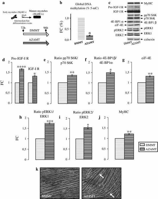
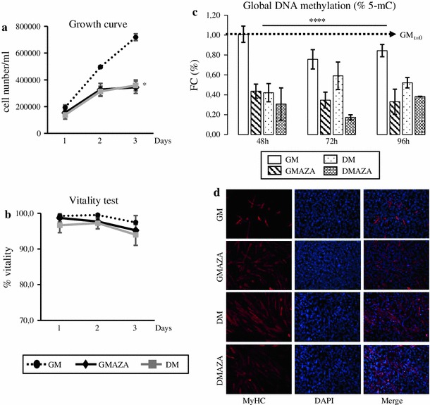
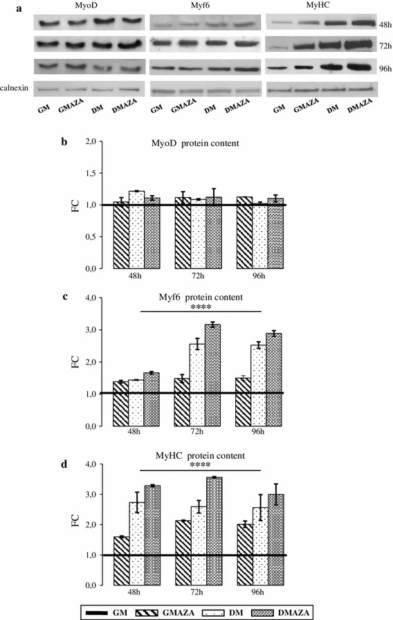
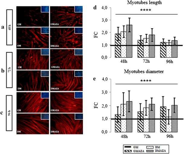
Skeletal muscle regeneration and hypertrophy are important adaptive responses to both physical activity and pathological stimuli. This research was performed to investigate DNA demethylation action on the late phase of muscle differentiation and early stage of hypertrophy. The epigenetic process involved in myogenesis was studied with the DNA-demethylating agent 5-azacytidine (AZA). We induced muscle differentiation in C2C12 mouse myoblasts in the presence of 5 μM AZA and growth or differentiation medium for 48, 72, and 96 h. To study a potential AZA hypertrophic effect, we stimulated 72 h differentiated myotubes with AZA for 24 h. Unstimulated cells were used as control. By western blot and immunofluorescence analysis, we examined AZA action on myogenic regulatory factors expression, hypertrophic signaling pathway and myotube morphology. During differentiation, protein levels of myogenic markers, Myf6 and Myosin Heavy Chain (MyHC), were higher in AZA stimulated cells compared to control. Myostatin and p21 analysis revealed morphological changes which reflect a tendency to hypertrophy in myotubes. In AZA stimulated neo formed myotubes, we observed that IGF-I pathway, kinases p70 S6, 4E-BP1, and ERK1/2 were activated. Furthermore, AZA treatment increased MyHC protein content in stimulated neo myotubes. Our work demonstrates that DNA demethylation could plays an important role in promoting the late phase of myogenesis, activating endocellular pathways involved in protein increment and stimulating the hypertrophic process.
骨骼肌再生和肥大是对身体活动和病理刺激的重要适应性反应。本研究旨在探讨DNA去甲基化对肌肉分化后期和肥大早期的作用。使用DNA去甲基化剂5-氮杂胞苷(AZA)研究了成肌过程中涉及的表观遗传过程。我们在存在5μM AZA以及生长或分化培养基的情况下诱导C2C12小鼠成肌细胞分化48、72和96小时。为了研究AZA的潜在肥大作用,我们用AZA刺激72小时分化的肌管24小时。未刺激的细胞用作对照。通过蛋白质印迹和免疫荧光分析,我们检测了AZA对肌源性调节因子表达、肥大信号通路和肌管形态的作用。在分化过程中,与对照相比AZA刺激的细胞中肌源性标志物Myf6和肌球蛋白重链(MyHC)的蛋白质水平更高。肌肉生长抑制素和p21分析揭示了形态学变化,这反映了肌管肥大的趋势。在AZA刺激的新形成的肌管中,我们观察到IGF-I途径、激酶p70 S6、4E-BP1和ERK1/2被激活。此外,AZA处理增加了刺激的新肌管中MyHC蛋白含量。我们的工作表明,DNA去甲基化在促进成肌后期、激活参与蛋白质增加的细胞内途径以及刺激肥大过程中可能发挥重要作用。