Labudda Kirsten, Kreisel Stefan, Beblo Thomas, Mertens Markus, Kurlandchikov Oleg, Bien Christian G, Driessen Martin, Woermann Friedrich G
Mara Hospital, Bethel Epilepsy Center, MRI Unit, Bielefeld, Germany ; Department of Psychiatry and Psychotherapy Bethel, Evangelisches Krankenhaus Bielefeld, Bielefeld, Germany.
Department of Psychiatry and Psychotherapy Bethel, Evangelisches Krankenhaus Bielefeld, Bielefeld, Germany.
PLoS One. 2013 Dec 18;8(12):e83677. doi: 10.1371/journal.pone.0083677. eCollection 2013.
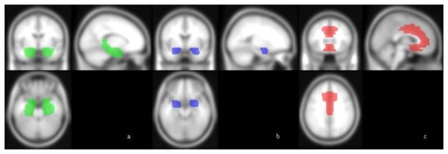
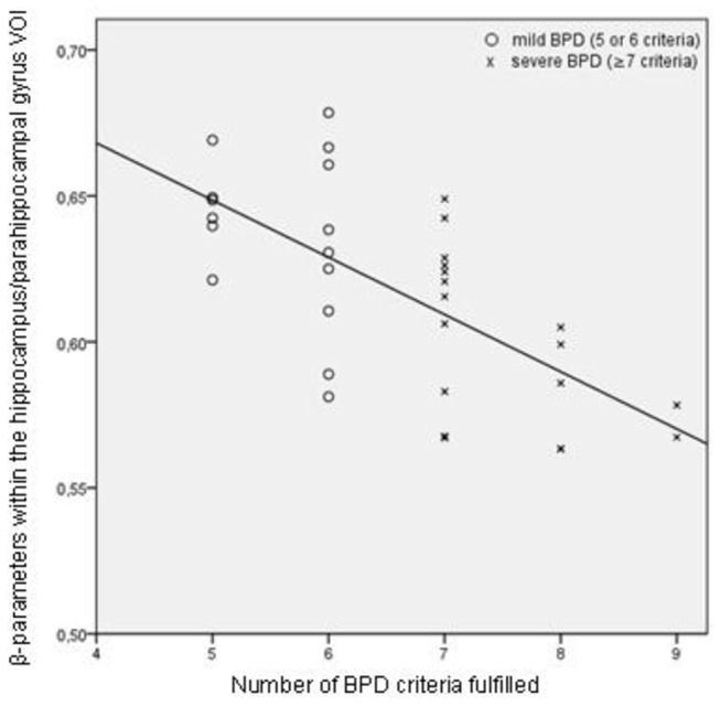
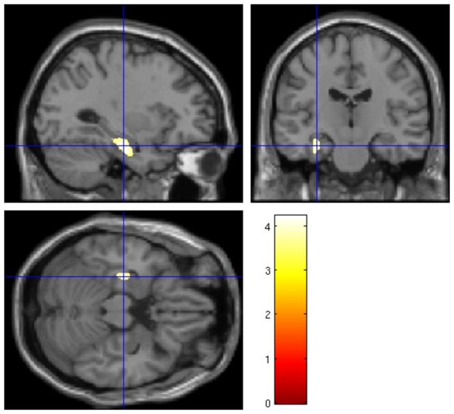
Results of MRI volumetry in Borderline Personality Disorder (BPD) are inconsistent. Some, but not all, studies reported decreased hippocampus, amygdala, and/or prefrontal volumes. In the current study, we used rater-independent voxel-based morphometry (VBM) in 33 female BPD patients and 33 healthy women. We measured gray matter (GM) volumes of the whole brain and of three volumes of interest (VOI), i.e., the hippocampus/parahippocampal gyrus, the amygdala and the anterior cingulate gyrus (ACC). Analyses were conducted using lifetime diagnoses of posttraumatic stress disorder (PTSD) and major depression (MD) as covariates. We used adversive childhood experiences and the numbers of BPD criteria (as an indicator of disorder severity) to investigate associations with GM volumes. We did not find volume differences between BPD patients and healthy subject, neither of the whole brain nor of the three VOIs, independent of presence or absence of comorbid PTSD and MD. We also did not find a relationship between childhood maltreatment and the patients' brain volumes. However, within the patient group, the number of BPD criteria fulfilled was inversely correlated with left hippocampal/parahippocampal volume (x=-32, y=-23, z=-18, k=496, t=5.08, p=.007). Consequently, mesiotemporal GM volumes do not seem to differentiate patients from healthy subjects, but might be associated with symptom severity within the BPD group.
边缘型人格障碍(BPD)的磁共振成像容积分析结果并不一致。部分(而非全部)研究报告称海马体、杏仁核和/或前额叶体积减小。在本研究中,我们对33名女性BPD患者和33名健康女性使用了独立于评分者的基于体素的形态测量法(VBM)。我们测量了全脑以及三个感兴趣区域(VOI),即海马体/海马旁回、杏仁核和前扣带回(ACC)的灰质(GM)体积。分析时将创伤后应激障碍(PTSD)和重度抑郁症(MD)的终生诊断作为协变量。我们使用童年不良经历和BPD标准数量(作为疾病严重程度的指标)来研究与GM体积的关联。我们未发现BPD患者与健康受试者之间在全脑或三个VOI的体积存在差异,无论是否存在共病PTSD和MD。我们也未发现童年虐待与患者脑体积之间存在关联。然而,在患者组中,满足的BPD标准数量与左侧海马体/海马旁回体积呈负相关(x = -32,y = -23,z = -18,k = 496,t = 5.08,p = 0.007)。因此,颞叶内侧GM体积似乎无法区分患者与健康受试者,但可能与BPD组内的症状严重程度相关。