Brain Research Institute, School of Medicine and Health Sciences, Monash University Malaysia, Petaling Jaya, 46150, Selangor, Malaysia.
Cell Tissue Res. 2014 Feb;355(2):409-23. doi: 10.1007/s00441-013-1765-9. Epub 2013 Dec 28.
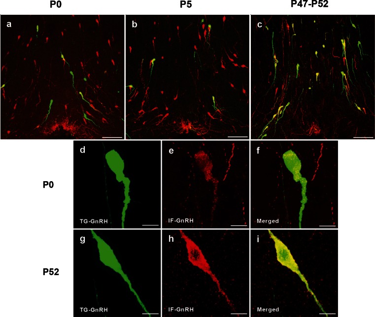
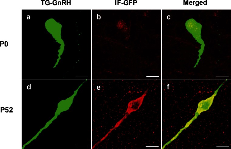
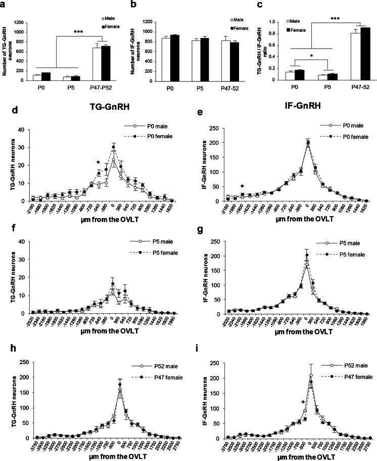
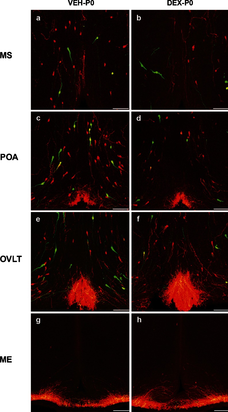
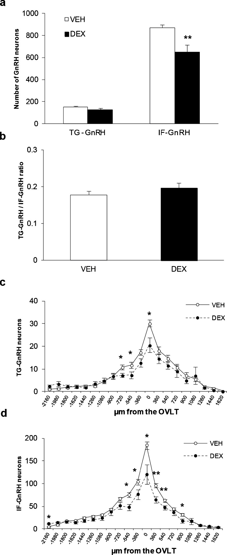
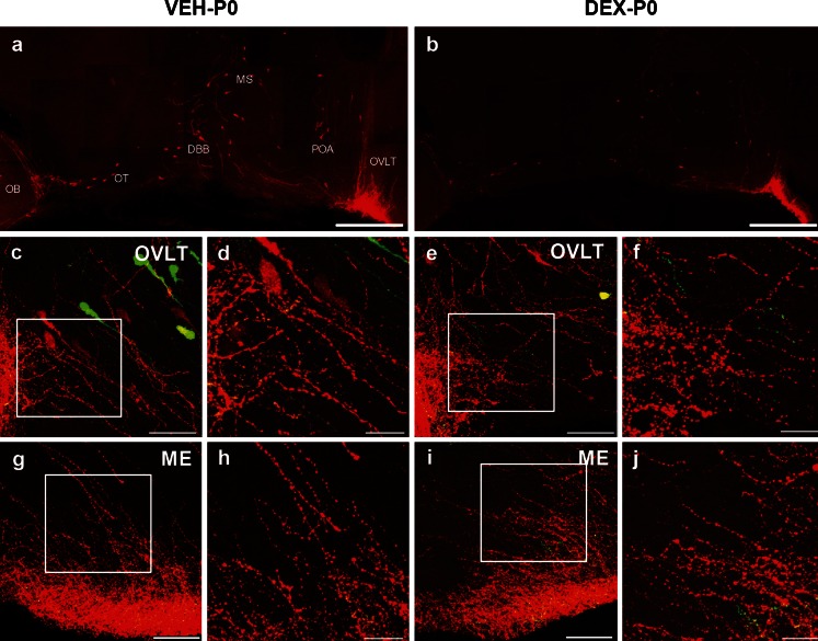
The migration of gonadotropin-releasing hormone (GnRH) neurons from the olfactory placode to the preoptic area (POA) from embryonic day 13 is important for successful reproduction during adulthood. Whether maternal glucocorticoid exposure alters GnRH neuronal morphology and number in the offspring is unknown. This study determines the effect of maternal dexamethasone (DEX) exposure on enhanced green fluorescent protein (EGFP) driven by GnRH promoter neurons (TG-GnRH) in transgenic rats dual-labelled with GnRH immunofluorescence (IF-GnRH). The TG-GnRH neurons were examined in intact male and female rats at different postnatal ages, as a marker for GnRH promoter activity. Pregnant females were subcutaneously injected with DEX (0.1 mg/kg) or vehicle daily during gestation days 13-20 to examine the number of GnRH neurons in P0 male offspring. The total number of TG-GnRH neurons and TG-GnRH/IF-GnRH neuronal ratio increased from P0 and P5 stages to P47-52 stages, suggesting temporal regulation of GnRH promoter activity during postnatal development in intact rats. In DEX-treated P0 males, the number of IF-GnRH neurons decreased within the medial septum, organum vasculosom of the lamina terminalis (OVLT) and anterior hypothalamus. The percentage of TG-GnRH neurons with branched dendritic structures decreased in the OVLT of DEX-P0 males. These results suggest that maternal DEX exposure affects the number and dendritic development of early postnatal GnRH neurons in the OVLT/POA, which may lead to altered reproductive functions in adults.
促性腺激素释放激素 (GnRH) 神经元从胚胎第 13 天从嗅基板迁移到视前区 (POA) 对于成年期的成功繁殖至关重要。母体糖皮质激素暴露是否会改变后代 GnRH 神经元的形态和数量尚不清楚。本研究确定了母体地塞米松 (DEX) 暴露对转基因大鼠中 GnRH 启动子驱动的增强型绿色荧光蛋白 (EGFP) (TG-GnRH) 的影响,这些大鼠用 GnRH 免疫荧光 (IF-GnRH) 双重标记。在不同的产后年龄,完整的雄性和雌性大鼠中检查了 TG-GnRH 神经元,作为 GnRH 启动子活性的标志物。在妊娠第 13-20 天期间,每天给怀孕的雌性大鼠皮下注射 DEX (0.1mg/kg) 或载体,以检查 P0 雄性后代中 GnRH 神经元的数量。从 P0 和 P5 阶段到 P47-52 阶段,TG-GnRH 神经元的总数和 TG-GnRH/IF-GnRH 神经元的比例增加,这表明 GnRH 启动子活性在完整大鼠的产后发育过程中存在时间调节。在 DEX 处理的 P0 雄性大鼠中,IF-GnRH 神经元的数量在内侧隔、终板血管器官 (OVLT) 和下丘脑前区减少。DEX-P0 雄性大鼠 OVLT 中具有分支树突结构的 TG-GnRH 神经元的百分比减少。这些结果表明,母体 DEX 暴露会影响 OVLT/POA 中早期产后 GnRH 神经元的数量和树突发育,这可能导致成年后生殖功能改变。