Rocher Crystal, Singla Dinender K
Biomolecular Science Center, Burnett School of Biomedical Sciences, College of Medicine, University of Central Florida, Orlando, Florida, United States of America.
PLoS One. 2013 Dec 20;8(12):e84009. doi: 10.1371/journal.pone.0084009. eCollection 2013.
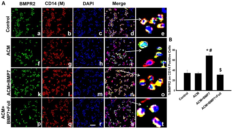
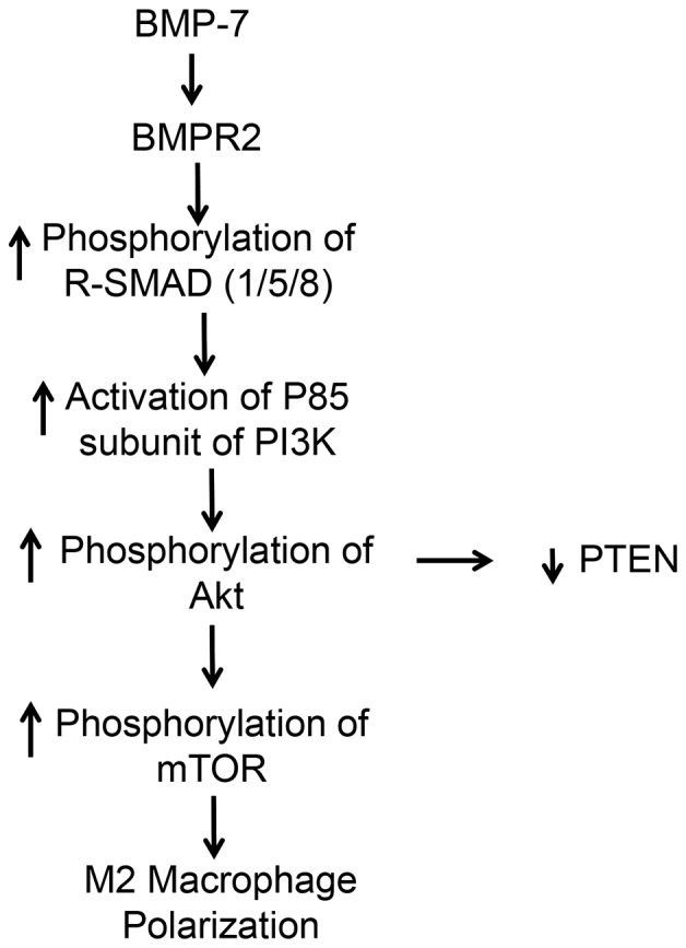
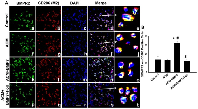
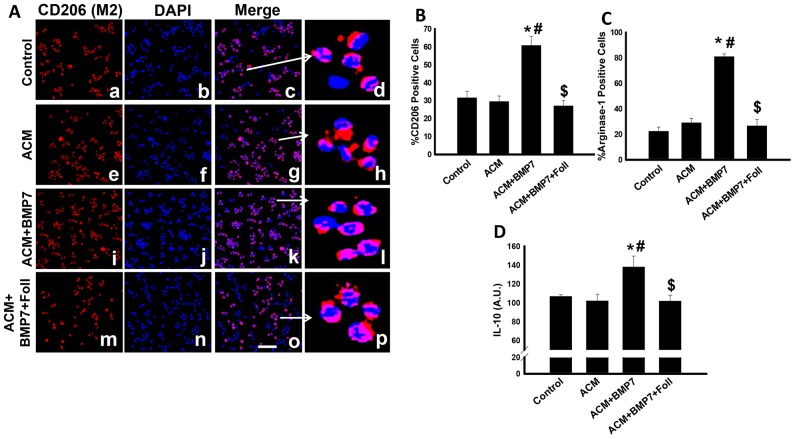
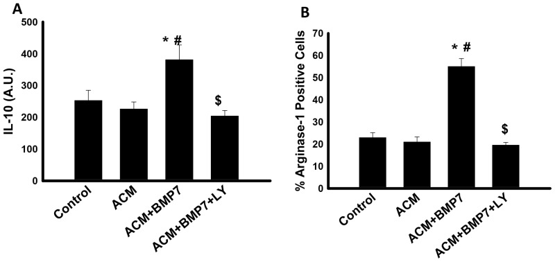
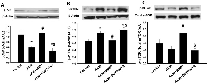
Previously we demonstrated that bone morphogenetic protein-7 (BMP-7) treatment polarizes monocytes into M2 macrophages and increases the expression of anti-inflammatory cytokines. Despite these findings, the mechanisms for the observed BMP-7 induced monocyte polarization into M2 macrophages are completely unknown. In this study, we demonstrate the mechanisms involved in the polarization of monocytes into M2 macrophages. Apoptotic conditioned media (ACM) was generated to mimic the stressed conditions, inducing monocyte polarization. Monocytes were treated with ACM along with BMP-7 and/or its inhibitor, follistatin, for 48 hours. Furthermore, an inhibitor of the PI3K pathway, LY-294002, was also studied. Our data show that BMP-7 induces polarization of monocytes into M2 macrophages while significantly increasing the expression of anti-inflammatory markers, arginase-1 and IL-10, and significantly (p<0.05) decreasing the expression of pro-inflammatory markers iNOS, IL-6, TNF-α and MCP-1; (p<0.05). Moreover, addition of the PI3K inhibitor, LY-294002, significantly (p<0.05) decreases upregulation of IL-10 and arginase-1, suggesting involvement of the PI3K pathway in M2 macrophage polarization. Next, following BMP-7 treatment, a significant (p<0.05) increase in p-SMAD1/5/8 and p-PI3K expression resulting in downstream activation of p-Akt and p-mTOR was observed. Furthermore, expression of p-PTEN, an inhibitor of the PI3K pathway, was significantly (p<0.05) increased in the ACM group. However, BMP-7 treatment inhibited its expression, suggesting involvement of the PI3K-Akt-mTOR pathway. In conclusion, we demonstrate that BMP-7 polarizes monocytes into M2 macrophages and enhances anti-inflammatory cytokine expression which is mediated by the activated SMAD-PI3K-Akt-mTOR pathway.
此前我们证明,骨形态发生蛋白-7(BMP-7)处理可使单核细胞极化为M2巨噬细胞,并增加抗炎细胞因子的表达。尽管有这些发现,但BMP-7诱导单核细胞极化为M2巨噬细胞的机制仍完全未知。在本研究中,我们阐述了单核细胞极化为M2巨噬细胞所涉及的机制。制备凋亡条件培养基(ACM)以模拟应激条件,诱导单核细胞极化。单核细胞用ACM以及BMP-7和/或其抑制剂卵泡抑素处理48小时。此外,还研究了PI3K途径的抑制剂LY-294002。我们的数据表明,BMP-7诱导单核细胞极化为M2巨噬细胞,同时显著增加抗炎标志物精氨酸酶-1和IL-10的表达,并显著(p<0.05)降低促炎标志物诱导型一氧化氮合酶(iNOS)、IL-6、肿瘤坏死因子-α(TNF-α)和单核细胞趋化蛋白-1(MCP-1)的表达;(p<0.05)。此外,添加PI3K抑制剂LY-294002显著(p<0.05)降低IL-10和精氨酸酶-1的上调,表明PI3K途径参与M2巨噬细胞极化。接下来,在BMP-7处理后,观察到p-SMAD1/5/8和p-PI3K表达显著(p<0.05)增加,导致下游p-Akt和p-mTOR激活。此外,PI3K途径的抑制剂p-PTEN的表达在ACM组中显著(p<0.05)增加。然而,BMP-7处理抑制了其表达,表明PI3K-Akt-mTOR途径参与其中。总之,我们证明BMP-7使单核细胞极化为M2巨噬细胞并增强抗炎细胞因子表达,这是由激活的SMAD-PI3K-Akt-mTOR途径介导的。