University of North Texas Health Science Center, North Texas Eye Research Institute, Department of Cell Biology and Anatomy, United States.
University of Alabama at Birmingham, Department of Visual Sciences, United States.
Cell Signal. 2014 Apr;26(4):665-672. doi: 10.1016/j.cellsig.2013.12.008. Epub 2013 Dec 27.
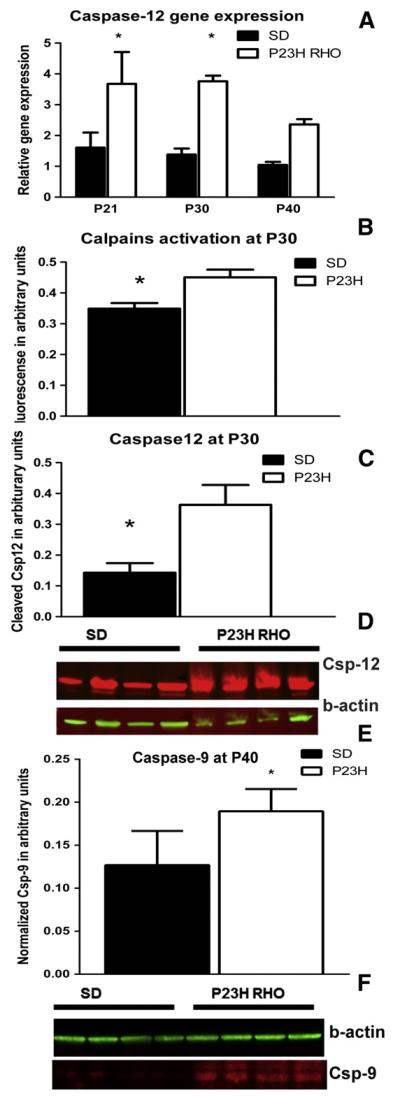
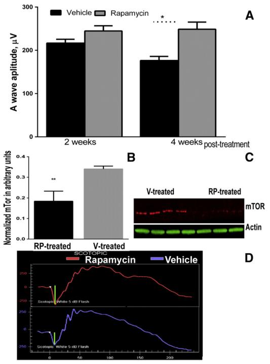
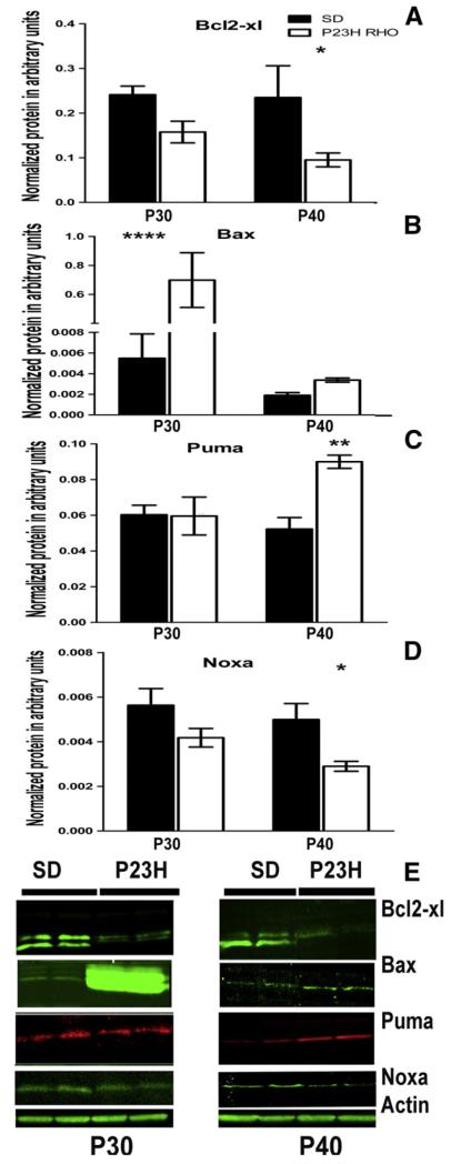
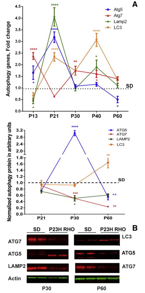
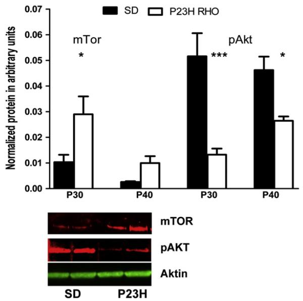
We previously reported activation of the unfolded protein response (UPR) in P23H rhodopsin (RHO) retinas with autosomal dominant retinitis pigmentosa (ADRP). Knowing that the UPR can trigger Ca(2+) release from the endoplasmic reticulum and regulate cellular signaling we examined the level of Ca(2+)-regulated proteins. We also looked for changes in the expression of Bcl2 family proteins, autophagy proteins and the mTOR/AKT pathways, as well as for the induction of mitochondria-associated apoptosis in the P23H RHO retina. Our data demonstrated that the elevation of calpain and caspase-12 activity was concomitantly observed with a decrease in the BCL2-XL/BAX ratio and an increase in mTor levels in the P23H-3 RHO retina suggesting a vulnerability of P23H RHO photoreceptors to apoptosis. The translocation of BAX to the mitochondria, as well as the release of cytochrome C and AIF into the cytosol supports this conclusion and indicates the involvement of mitochondria-induced apoptosis in the progression of ADRP. The level of autophagy proteins in general was found to be decreased in the P21-P30 P23H RHO retina. Injections of rapamycin, however, protected the P23H RHO rod photoreceptors from experiencing physiological decline. Despite this fact, the downregulation of mTOR did not alter the level of autophagy proteins. Our results imply that in addition to activation of the UPR during ADRP progression, photoreceptors also experience alterations in major proapoptotic pathways.
我们之前曾报道过常染色体显性遗传视网膜色素变性(ADRP)中 P23H 视紫红质(RHO)视网膜中未折叠蛋白反应(UPR)的激活。由于 UPR 可以触发内质网中 Ca2+的释放并调节细胞信号,我们检查了 Ca2+调节蛋白的水平。我们还观察了 Bcl2 家族蛋白、自噬蛋白和 mTOR/AKT 途径的表达变化,以及 P23H RHO 视网膜中线粒体相关凋亡的诱导。我们的数据表明,在 P23H-3 RHO 视网膜中,钙蛋白酶和半胱天冬酶-12 活性的升高与 BCL2-XL/BAX 比值的降低和 mTor 水平的升高同时观察到,提示 P23H RHO 光感受器对凋亡的敏感性增加。BAX 向线粒体的易位,以及细胞色素 C 和 AIF 向细胞质的释放支持这一结论,并表明线粒体诱导的凋亡参与了 ADRP 的进展。在 P21-P30 P23H RHO 视网膜中,一般发现自噬蛋白的水平降低。然而,雷帕霉素的注射保护了 P23H RHO 视杆细胞免受生理衰退的影响。尽管如此,mTOR 的下调并没有改变自噬蛋白的水平。我们的结果表明,除了 ADRP 进展过程中 UPR 的激活之外,光感受器还经历了主要促凋亡途径的改变。