Khorsand Afshin, Paknejad Mojgan, Yaghobee Siamak, Ghahroudi Amir Alireza Rasouli, Bashizadefakhar Hourieh, Khatami Masoomeh, Shirazi Mohsen
Department of Periodontology, Tehran University of Medical Sciences, Tehran, Iran.
Department of Periodontology and Dental Implant Research Center, Tehran University of Medical Sciences, Tehran, Iran.
Dent Res J (Isfahan). 2013 Nov;10(6):744-51.
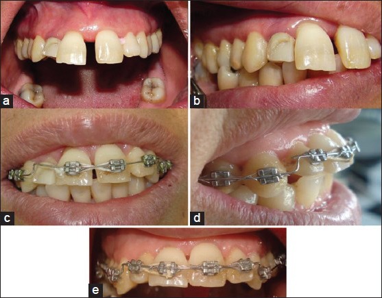
The success of combined periodontal and orthodontic approach in the treatment of aggressive periodontitis patients with the pathologic extruded anterior teeth is a main concern and stability of the treatment results is an important factor to evaluate the treatment. The present study investigated the periodontal parameters at the end of the orthodontic treatment in patients with the aggressive periodontitis.
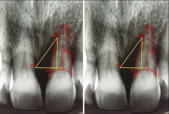
Eight patients with an aggressive periodontal disease, extruded maxillary incisors, infrabony defects and probing depth of ≥5 mm were enrolled in this clinical trial (before, after). After periodontal therapy, orthodontic treatment was carried out for intrusion and alignment of teeth. Plaque index (PI), probing pocket depth (PPD), distance between incisal edge and interdental papilla, root length (RL), and defect dimensions (depth and width) were examined at the end of treatment and three as well as 6 months afterward. The data were subjected to repeated measure ANOVA test. P < 0.05 was considered as significant.
There was statistically significant decrease in PPD, PI, and depth of the defects during T0, T3 and T6 (P < 0.05). No significant differences were observed in the RL and distance between incisal edge and interdental papilla (P = 0.95). Furthermore, width of the defects demonstrated significant decrease up to T3 (P = 0.042) while no significant changes from 3 months to 6 months were noted (P = 0.59).
The results showed that combined periodontal and orthodontic approach would be a successful treatment with acceptable stability in the case of regular follow-up visits and controlled oral hygiene habits.
牙周治疗与正畸联合治疗侵袭性牙周炎伴病理性前牙前突患者的疗效是主要关注点,治疗结果的稳定性是评估该治疗的重要因素。本研究调查了侵袭性牙周炎患者正畸治疗结束时的牙周参数。
本临床试验纳入8例患有侵袭性牙周病、上颌切牙前突、骨下袋且探诊深度≥5 mm的患者(治疗前、治疗后)。牙周治疗后,进行正畸治疗以压低并排齐牙齿。在治疗结束时以及之后3个月和6个月检查菌斑指数(PI)、探诊袋深度(PPD)、切缘与龈乳头之间的距离、牙根长度(RL)以及骨缺损尺寸(深度和宽度)。数据采用重复测量方差分析检验。P < 0.05被认为具有统计学意义。
在T0、T3和T6期间,PPD、PI和骨缺损深度有统计学意义的下降(P < 0.05)。RL以及切缘与龈乳头之间的距离未观察到显著差异(P = 0.95)。此外,骨缺损宽度在T3时显示出显著下降(P = 0.042),而从3个月到6个月未观察到显著变化(P = 0.59)。
结果表明,在定期随访和良好口腔卫生习惯控制的情况下,牙周治疗与正畸联合治疗是一种成功且稳定性可接受的治疗方法。