Institute for Lung Health, Department of Infection, Immunity and Inflammation, University of Leicester, Leicester, United Kingdom.
Division of Respiratory Medicine, Centre for Respiratory Research and Nottingham Respiratory Biomedical Research Unit, University of Nottingham, Nottingham, United Kingdom.
PLoS One. 2013 Dec 31;8(12):e85244. doi: 10.1371/journal.pone.0085244. eCollection 2013.
Idiopathic pulmonary fibrosis (IPF) is a common, progressive and invariably lethal interstitial lung disease with no effective therapy. We hypothesised that K(Ca)3.1 K(+) channel-dependent cell processes contribute to IPF pathophysiology.
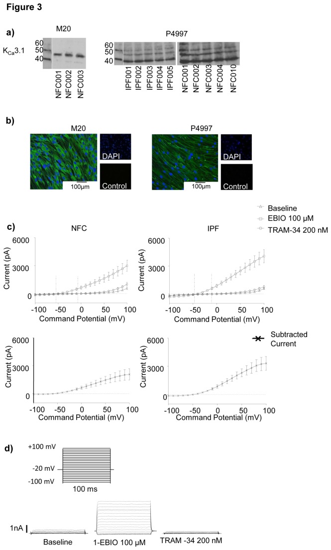
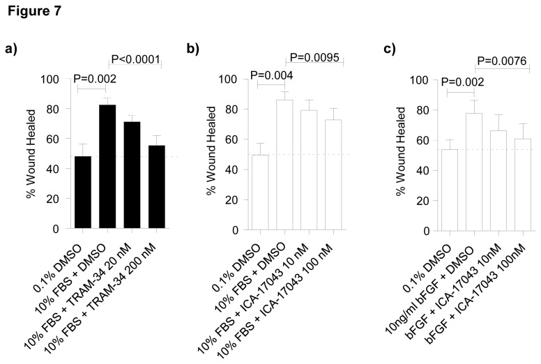
K(Ca)3.1 expression in primary human lung myofibroblasts was examined using RT-PCR, western blot, immunofluorescence and patch-clamp electrophysiology. The role of K(Ca)3.1 channels in myofibroblast proliferation, wound healing, collagen secretion and contraction was examined using two specific and distinct K(Ca)3.1 blockers (TRAM-34 and ICA-17043 [Senicapoc]).
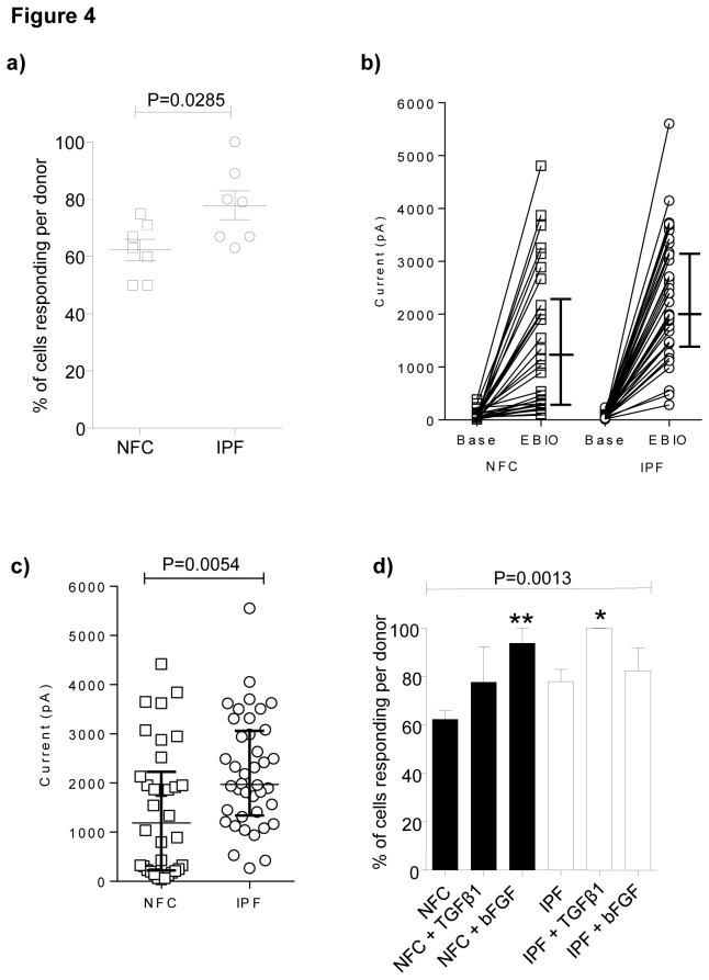
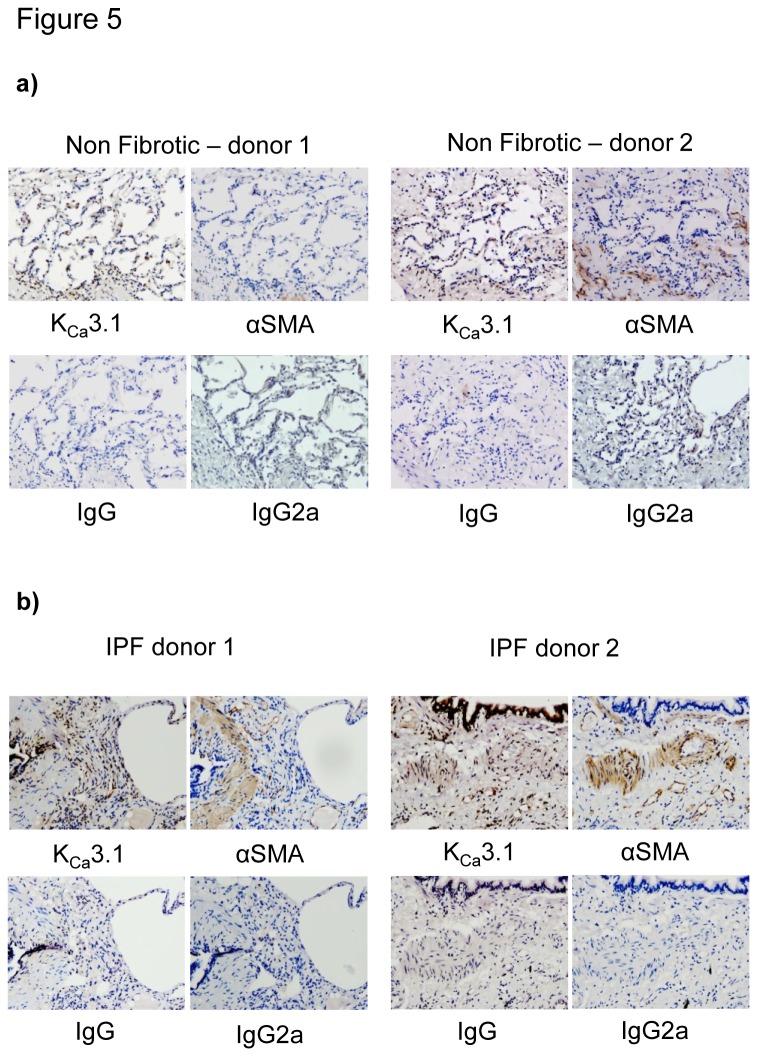
Both healthy non fibrotic control and IPF-derived human lung myofibroblasts expressed K(Ca)3.1 channel mRNA and protein. K(Ca)3.1 ion currents were elicited more frequently and were larger in IPF-derived myofibroblasts compared to controls. K(Ca)3.1 currents were increased in myofibroblasts by TGFβ1 and basic FGF. K(Ca)3.1 was expressed strongly in IPF tissue. K(Ca)3.1 pharmacological blockade attenuated human myofibroblast proliferation, wound healing, collagen secretion and contractility in vitro, and this was associated with inhibition of TGFβ1-dependent increases in intracellular free Ca(2+).
K(Ca)3.1 activity promotes pro-fibrotic human lung myofibroblast function. Blocking K(Ca)3.1 may offer a novel approach to treating IPF with the potential for rapid translation to the clinic.
特发性肺纤维化(IPF)是一种常见的、进行性的、致命性的间质性肺疾病,目前尚无有效的治疗方法。我们假设 K(Ca)3.1 钾通道依赖性细胞过程有助于 IPF 的病理生理学。
使用 RT-PCR、western blot、免疫荧光和膜片钳电生理学检测原代人肺成纤维细胞中 K(Ca)3.1 的表达。使用两种特异性和不同的 K(Ca)3.1 阻滞剂(TRAM-34 和 ICA-17043[Senicapoc])研究 K(Ca)3.1 通道在成纤维细胞增殖、伤口愈合、胶原分泌和收缩中的作用。
健康非纤维化对照和 IPF 来源的人肺成纤维细胞均表达 K(Ca)3.1 通道 mRNA 和蛋白。与对照组相比,IPF 来源的成纤维细胞中 K(Ca)3.1 离子电流的激发频率更高,幅度更大。TGFβ1 和碱性成纤维细胞生长因子可增加成纤维细胞中的 K(Ca)3.1 电流。K(Ca)3.1 在 IPF 组织中表达强烈。K(Ca)3.1 药理学阻断可减弱体外人成纤维细胞增殖、伤口愈合、胶原分泌和收缩性,这与抑制 TGFβ1 依赖性细胞内游离 Ca(2+)增加有关。
K(Ca)3.1 活性促进了促纤维化的人肺成纤维细胞功能。阻断 K(Ca)3.1 可能为治疗 IPF 提供一种新方法,具有快速转化为临床应用的潜力。